Image Processing Reference
In-Depth Information
voxels of the LR image. To a
developed to estimate the HR
image degradation process [
where only one LR image is
means upsampling was propo
and total variation are used
process. While these metho
information from multiple im
Longitudinal studies are
structural and functional de
tiple times, such as at birth
contrast in neonatal image
follow-up images for guidi
The reason is that, the maj
fine-tuned after birth [9]. Fi
affine alignment. Despite t
remain consistent longitudi
subject share the identical b
tion than those images from
In this paper, we propose
natal image from a neonatal
Specifically, since the follo
contrast, they are ideal for
images (Fig. 1). We first u
structures in high-resolution
high-resolution reconstructi
fold: 1) We learn longitudin
and total variation regulariza
citly model the image degra
proposed method will be eva
other state-of-the-art method
address this issue, super-resolution (SR) techniques have b
R image from one or more LR input images by reverting
[1, 5]. Many existing approaches focus on single-frame
s available to recover the HR image. For example, non-lo
osed for HR image reconstruction in [6]. In [7], both low-r
d to regularize the otherwise ill-posed image reconstruct
ods have been shown to be effective, using complement
mages might help improve reconstruction accuracy.
e widely employed to investigate the dynamic early br
evelopments. In this setting, a subject is scanned for m
and 2 years of age. To address the challenges of low tis
es, recent studies have proposed to use their longitudi
ing the image processing such as tissue segmentation
or brain gyrification is established before birth while o
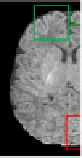
ig. 1 shows a neonatal image and its 2-year-old image a
the differences in image contrast, brain structural patte
inally. Meanwhile, since the longitudinal images of a sa
brain anatomy, they could be better matched after regis
m different subjects.
e a novel super-resolution method for recovering a HR n
l LR image using its longitudinal follow-up image as a pr
ow-up images typically have higher resolution and tis
guiding the resolution enhancement of the neonatal br
se a non-local approach to learn the spatial relationship
n longitudinal images and then apply this information to
ion of the neonatal image. Our main contribution is th
nal voxel relationship as a prior; 2) We integrate low-r
ation for effective estimation of the HR image; 3) We ex
adation processes involving blurring and downsampling. T
aluated using a group of neonatal images and compared w
ds.
been
g the
SR,
ocal
rank
tion
tary
rain
mul-
ssue
inal
[8].
only
after
erns
ame
stra-
neo-
rior.
ssue
rain
p of
the
hree
rank
xpli-
The
with
onate (left) and its follow-up at 2 years of age (right). The 2-y
the neonatal image using affine alignment. Two brain regi
re zoomed up for close comparison.
Fig. 1.
T1 MR images of a neo
old image was registered to
marked with green and red wer
year-
ions
2
Method
We propose a novel method
we briefly introduce the s
d for neonatal image super-resolution reconstruction. Fi
super-resolution problem. Next, we put emphasis on
irst,
the


Search WWH ::

Custom Search