Chemistry Reference
In-Depth Information
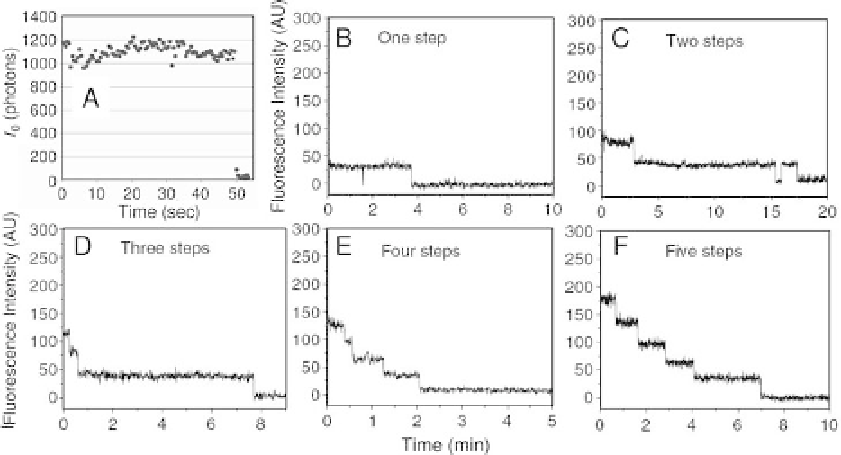
Figure 3.4 Discrete photobleaching of small numbers of
fluorophores. A, single bifunctional rhodaminemolecule lasts 50 s
under illumination in oxygen-depleted buffer (from ref. [27]). B-F,
intensity traces for bleaching of one to five Cy3 pRNAmoleculae in
a viral packaging motor (from Ref. [28]).
photobleaching in one step, indicate that the emission is from an individual
probe.
When two or more probes occupy the same picture element, the intensity is higher
(not always by an integer multiple, due to quenching by energy transfer) and the
photobleaching occurs in multiple steps (Figure 3.4B
-
F). For oligomeric complexes,
it is possible to estimate the number of components from the number of bleaching
steps [28
-
30]. Detection of individual
fluorescent molecules or small groups
has become straightforward with commercially available or readily constructed
instrumentation. The research effort now has shifted toward obtaining useful,
mechanistically relevant, biological information.
3.2.2
Sub-Diffraction Localization of Fluorescent Molecules
Single molecule imaging has overcome the limit of spatial resolution in optical
microscopy that was formerly thought to be insurmountable. The location of
individual
fluorophores can be determined with an uncertainty of a few nan-
ometers, a range readily suited to studying the 8
-
36-nm stepping motions of
molecular motors. In a
fluorescence microscope, the emission from a point source
is broadened at the detector due to diffraction and aberrations in the microscope
optics. The point spread function (PSF) of the microscope is the apparent spatial
distribution of the origin of the photons emitted from a source with essentially
in
nitesimal size, such as an individual
fluorescent probe. Generally, the PSF is a