Chemistry Reference
In-Depth Information
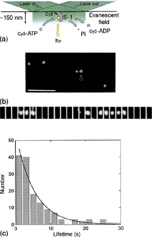
Figure 2.2 Single molecule imaging of the
enzymatic (ATPase) reaction by myosin [2].
(a) TIRFM for the observation of individual ATP
turnovers by a single myosin molecule.
(b) Fluorescence images of single myosin
molecules labeled with Cy-5. Scale bar
of fluorescence from a Cy3 nucleotide (ATP
or ADP) coming in (down arrows) and out
(upper arrows) of focus by associating and
dissociating with a myosin molecule. Images
were taken every 3 s. (c) A histogram of
lifetimes of a Cy3
¼
5mm
nucleotide bound to
myosin. Solid line shows an exponential fit
to the data.
-
(top). ATP turnovers by a single myosin
molecule; the lower panel shows typical images
associated with myosin bound to a surface, the Brownian motion ceases and the
labeled nucleotide can be observed as a clear
fluorescent spot. Thus, association and
dissociation of Cy3-nucleotides (ATP or ADP) can be observed by monitoring the
flickering of
fluorescent spots. Because the af
nity of ATP for muscle myosin is
10
5
-fold higher than that of ADP, the nucleotide coming into focus when associat-
ing with myosin should be Cy3-ATP and that coming out of focus when dissociating
from the myosin should be Cy3-ADP after hydrolysis. Thus, the association and
dissociation cycle of Cy3
-
nucleotides with myosin should be directly coupled to the