Chemistry Reference
In-Depth Information
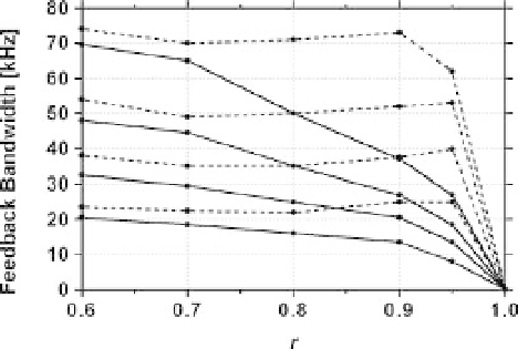
Figure 12.3 Effect of dynamic PID control on the feedback
bandwidth of tappingmode AFM. These data were experimentally
obtained. Solid lines: a conventional PID controller was used;
broken lines: a dynamic PID controller was used. The solid-line
curves and the dotted-line curves are aligned from top to bottom
according to the ratios 2A
0
/h
0
¼
5, 2, 1, and 0.5. A z-scanner with a
bandwidth of 150 kHz was used.
sample and substratum preparation methods necessary to image biomolecular
processes. These efforts have steadily improved the quality of captured images,
the imaging rate, and the magnitude of the tip-sample interaction force. Imaging
experiments performed most recently captured dynamic protein
-
protein inter-
actions on video with an imaging performance capable of revealing molecular
mechanisms. We describe below some imaging experiments in chronological
order.
In 2001, we reported the
first image data successively captured at 80ms/frame.
The sample was myosin V weakly attached to a mica surface in a solution containing
ATP [2]. Although the images were noisy, the swinging-lever-like movement was
visible. Since the ATPase rate of myosin V in the absence of ATP is low, this
movement was not observed repeatedly, which made interpretation of the data
inconclusive. Soon after, we attempted to observe the gliding movement of actin
filaments on a myosin V-coated surface. However, actin
filaments did not appear in
the scanning region. The reason was that actin
filaments were easily detached from
anchored myosin V by the scanning cantilever tip. In 2002, we developed a method
to combine UV-
ash photolysis of caged-compounds with high-speed AFM. A
strong UV-
ash bent a cantilever signi
cantly, resulting in a strong impact between
the tip and the substratum and hence damaging the tip. Therefore, many
flashes of
attenuated UV were applied while the sample stage was being scanned in y-return
after its slight withdrawal from the cantilever in the z-direction. We applied this
method to observe conformational changes in myosin V that occurred synchro-
nously with UV irradiation. Because of the synchronicity, the observed changes can
be interpreted as those really induced by ATP binding. In fact, immediately after UV
irradiation, the head portion bent around the head
-
neck junction and returned to its
original straight form (Figure 12.4a). This technique was also applied to the