Chemistry Reference
In-Depth Information
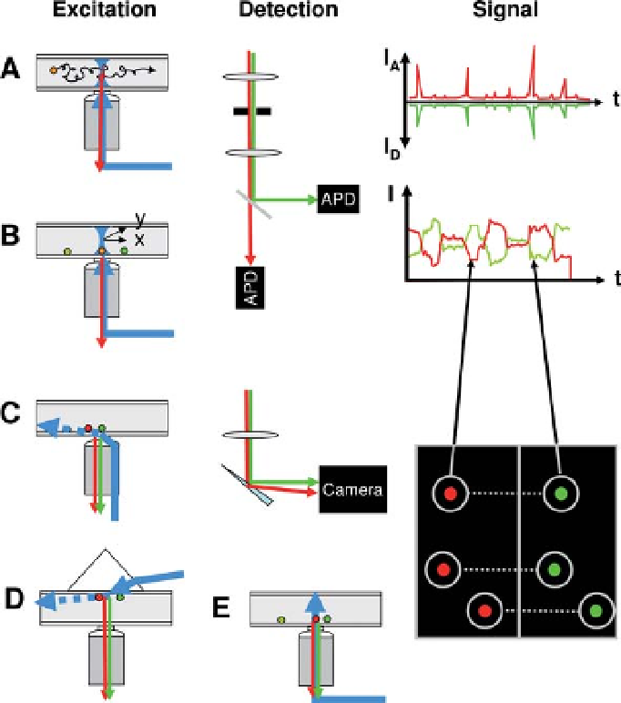
Figure 9.4 Experimental geometries used in
single-molecule fluorescence spectroscopy. Two
main types of geometries can be used for single-
molecule fluorescence spectroscopy: confocal
and wide-field. In the confocal geometry (A, B), a
collimated laser beam is sent into the back focal
plane of a high numerical aperture objective lens,
which focuses the excitation light into a
diffraction limited volume (or point spread
function
(but typically less than a few ms), as indicated
schematically on the right-hand side (RHS).
Immobile molecules (B) will first need to be
localized using a scanning device (indicated as
two perpendicular arrows x and y), before
recording can commence. Typical time traces are
comprised of one or more fluctuating intensity
levels until themolecule eventually bleaches after
a few seconds, as indicated schematically on the
RHS. The wide-field geometry (C
PSF) in the sample. Fluorescence
emitted by molecules present in this volume is
collected by the same objective and transmitted
through dichroic mirrors, lenses and color filters
to one or several point detectors (avalanche
photodiodes
E) can be used
in two different modes: (C, D) total internal
reflection (TIR) or (E) epifluorescence. In TIR, a
laser beam is shaped in such a way that a
collimated beam reaches the glass
-
-
buffer
-
sin
1
(n
buffer
/
n
glass
), where n designates the index of
refraction. This creates an evanescent wave
(decay length of a few 100 nm) in the sample
(dashed arrow), which only excites the
fluorescence of molecules in the vicinity of the
surface, resulting in very low background. TIR
APD). An important aspect of this
geometry is the presence of a pinhole in the
detection path, whose size is chosen such as to
let only light originating from the region of the
excitation PSF reach the detectors. Freely-
diffusing molecules (A) will yield signals
comprised of bursts of various size and duration
interface at a critical angle
q¼
-