Environmental Engineering Reference
In-Depth Information
photography are in darker shades, whereas eleva-
tions are in lighter ones. The main advantage of
using this method is that it enables the study of
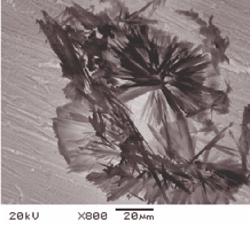
sample morphology; Figure 3.5 shows SEM images
of two clays (
SEM. The details of particle texture, as well as their
crystalline structure, can be studied in detail with this
technique. However, sample preparation is highly
complex, including granulometric separation, ionic
and molecular saturation, dilutions, dispersions,
dehydrations, impregnations, inclusion in resin,
drying, sectioning, and deposition on grids (Elsass
et al.
2008). Basically, two forms of sample prepara-
tion are possible: ultrathin sectioning and deposits.
In TEM, parallel electron beams pass through a
set of objective lenses and illuminate the sample, the
beams then spread out owing to interaction with it.
The primary image approximates the inverse Fourier
transform of the diffraction pattern and is subse-
quently magnifi ed by additional lenses to form the
fi nal image.
Using both SEM and TEM, low- or high-resolu-
tion cameras capture digital images, which are sub-
sequently treated using computer programs. This
procedure assures a high turnaround of image analy-
sis, so that information is obtained quickly, such as
particle dimension, structure, and interlayer spaces.
In Fig. 3.6, TEM micrographs of clays from sub-
tropical soils are presented where the morphology of
the clay mineral particles inside the resin can be seen
as well as individual particles.
The chemical composition of particles can also be
obtained using X-ray emission electronic microscopy
done in association with TEM and SEM. In both
techniques, one region of the image representing
many or only one particle can be selected by micro-
sound, where operating conditions, such as X-ray
intensity and observing time, are also controlled
(Elsass & Flores-Velez 1999). The image and elemen-
tal composition are obtained and analyzed simulta-
neously, and computer software further enables the
relation between structural and absorbed elements to
be explored (Dur
et al.
2004). From a practical view-
point, this information is valuable in studies of pol-
lution and pollutant transport (Citeau
et al.
2006).
m), illite and smectite, from fl uvial
sediment in a watershed in southern Brazil.
TEM observations are used for individual mineral
particle and ionic species associated with fi ne frac-
tions in sediments. Smaller particle sizes can be
studied with TEM owing to its better resolution than
<
2
μ
3.4 Nuclear magnetic resonance
3.4.1 Basic theory of nuclear magnetic
resonance
Fig. 3.5
Two clay mineral images obtained by scanning
electron microscopy from the fi ne clay fraction of sediments in
a watershed, southern Brazil (upper, illite; bottom, smectite).
Photographs obtained by a JEOL
®
SEM apparatus.
From Bortoluzzi
et al.
(2006).
Although most of the chemical properties of the
atoms are related only to the electrons that surround
the nucleus, there are some characteristics of the