Biology Reference
In-Depth Information
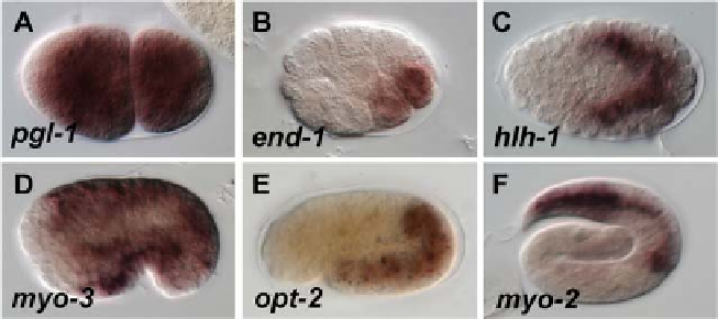
Fig. 5
Antisense RNA probes detect endogenous mRNAs consistent with published reports.
(A) Maternal transcripts of pgl-1 at the two-cell stage (
Kawasaki et al., 1998
). (B) Expression of end-1
in the E daughter cells (
Zhu et al., 1997
). (C) Expression of hlh-1 in muscle precursors (
Krause et al.,
1990
). (D) Activation of myo-3 in body muscle cells (
Okkema et al., 1993
). (E) Expression of opt-2 in
intestine cells (
Nehrke, 2003
). (F) Expression of myo-2 in pharynx muscle cells (
Okkema et al., 1993
).
Anterior is left, and dorsal is up. Embryos are approximately 50
m
m long. (For color version of this figure,
the reader is referred to the web version of this topic.)
index_e.html
)
, a platform that together costs approximately $1500, and which is
also suitable for fluorescence microscopy. It is also recommended that many
animals (
>
25) of specific stages be examined for staining, and that the number
of animals with good staining be quantified. Under optimal conditions, we rou-
tinely observe that at least 75%, and usually greater than 80%, of embryos will
show detectable signal (
Lin et al., 2009
). The sensitivity of the approach has been
confirmed by quantification of transcripts by single molecule detection (
Raj et al.,
2010
). Quantification of endogenous transcripts of zygotic genes expressed in
endoderm specification has shown that there are at most some 400 transcripts of
end-3 in the early E lineage (
Raj et al., 2010
). We have observed very strong
expression of end-3 in the E cell of early embryos (
Fig. 5B
)(
Maduro et al., 2007
),
suggesting that this procedure is sensitive enough to detect several hundred
transcripts. Given that the signals observed for end-3 are fairly strong, it is likely
that smaller numbers of transcripts (e.g., around 100) could be detected with this
approach.
X. Summary
The detection of mRNA in situ provides a rapid means by which to determine the
expression pattern of endogenous genes. A timetable is provided to assist in planning
(
Fig. 6
). We have described a protocol that, in our hands, results in reproducible
staining of endogenous mRNAs with a lower limit of at most several hundred

Search WWH ::

Custom Search