Biology Reference
In-Depth Information
limitations and advantages of each strategy will dictate which technique is best for a
given application.
G. Marking Extrachromosomal Arrays to Probe Gene Regulation
The interaction of the E. coli LacI protein with lacO lactose operator sequences
was exploited as a method for marking chromosomes in yeast (
Belmont and Straight,
1998
) and has been used as a marker for transgenes in C. elegans as well (
Gonzalez-
Serricchio and Sternberg, 2006
). Use of the LacI/LacO systems has also been used to
label extrachromosomal arrays to study gene regulation (
Fig. 3A
). In such experi-
ments, a GFP-tagged endogenous transcription factor is expressed in the presence of
an extrachromosomal array that carries a promoter that contains its target cis-
regulatory sites. The factor will interact with the many copies of the target promoter
in the array, producing a subnuclear spot. LacI tagged with a different marker can
label lacO sequences in the same target array, allowing an independent means by
which to verify interaction of the GFP-tagged factor with the array (
Carmi et al.,
1998
). Researchers have also used the GFP::LacI/LacO system to demonstrate that
transgenes move to different locations in the nucleus depending on whether they are
active or inactive in a given cell or tissue (
Meister et al., 2010
).
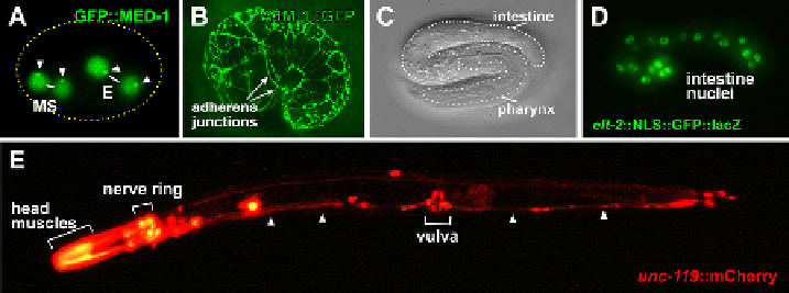
Fig. 3
Examples of types of transgenes and their expression patterns. (A) Expression of a chromosom-
ally-integrated med-1::GFP::MED-1 translational reporter in the early embryo, showing nuclear GFP
expression in the daughters of the blastomeres MS and E (
Maduro et al., 2002
). Due to the presence of a
separate extrachromosomal array carrying a transcriptional lacZ reporter for the MED-1 target gene end-3,
the GFP::MED-1 localizes to subnuclear spots representing the extrachromosomal array (arrowheads) in
each nucleus. (B) Expression of a translational fusion of the adherens junction marker ajm-1 in mid-
embryogenesis. GFP becomes localized to adherens junctions, giving an outline of epidermal cells
(
Koppen et al., 2001
). (C) DIC image of a late embryo, just prior to hatching, with the pharynx and intestine
indicated. (D) Expression of an elt-2::NLS::YFP::lacZ reporter transgene in the same embryo as in (C)
localized to intestinal nuclei (and excluded from nucleoli). (E) A C. elegans adult hermaphrodite showing
expression of an unc-119::mCherry transcriptional reporter throughout the nervous system (including the
nerve ring, neurons around the vulva, and the ventral nerve cord indicated by arrowheads) and in head muscles
(Maduro and Pilgrim, 1995). The head muscle expression has been overexposed. Anterior is to the left. A
C. elegans embryo is approximately 50
m
m long, while adults are approximately 1mm long. (See color plate.)
Search WWH ::

Custom Search