Biology Reference
In-Depth Information
Indo
50
1.5
Elec
EGTA
1
40
0.5
30
0
0
10
Time (s)
Elec
20
10
0
−
10
−
5
0
5
10
15
Time (s)
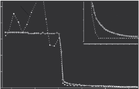
Fig. 3
Comparison of response time of a Ca
2
þ
-selective minielectrode and indo-1. Free [Ca
2
þ
] was
measured in a suspension of permeabilized rabbit ventricular myocytes. Ca
2
þ
uptake into sarcoplasmic
reticulum and mitochondria was inhibited with thapsigargin and ruthenium red, respectively. The initial
free [Ca
2
þ
] was 32
m
Mwhich is near saturation for indo-1, resulting in a very noisy trace. That is because
a small change in fluorescence ratio corresponds to a large in [Ca
2
þ
] at this level (see
Fig. 1
). At the
arrow, 2 mMEGTAwas added to the cell suspension to lower free [Ca
2
þ
]. Both the electrode and indo-1
signal were more than 90% complete in 1 s. The inset shows the response of indo-1 and Ca
2
þ
electrode at
low [Ca
2
þ
]. Notice that the indo-1 signal was 100% complete in 2 s (and actually undershoot slightly)
while the final completion of the electrode response was slower.
apparently slower electrode response may result from inhomogeneities rather than
a slower electrode response per se. This is illustrated in
Fig. 4
, where free [Ca
2
þ
]
was monitored with Ca
2
þ
electrode and indo-1 simultaneously in a myocyte
suspension in the absence and presence of oxalate.
In
Fig. 4
A, Ca
2
þ
addition to the cells causes a rapid increase in [Ca
2
þ
], which is
subsequently sequestered by the sarcoplasmic reticulum (SR). In the absence of
10 mM oxalate (
Fig. 4
A), the electrode response appears to be slower than the
corresponding indo-1 signal. However, when oxalate is subsequently added to the
cell suspension (
Fig. 4
B), the measured change in free [Ca
2
þ
] after a Ca
2
þ
addition
is similar for indo-1 and the Ca
2
þ
electrode. It should be noted that oxalate not
only bu
ers the free [Ca
2
þ
], but also increases the Ca
2
þ
uptake rate in the SR, and
thereby the removal of Ca
2
þ
from the cell suspension. Thus, despite inducing a
faster rate of change in free [Ca
2
þ
], oxalate eliminates the di
V
erence between Ca
2
þ
electrode and indo-1 signal by eliminating spatial inhomogeneities in free [Ca
2
þ
]in
the myocyte suspension. Indeed, indo-1 is expected to be less sensitive to spatial
inhomogeneities as it di
V
uses into the permeabilized cells and binds to cellular
proteins (
Hove-Madsen and Bers, 1992, 1993b
). In contrast, the Ca
2
þ
electrode
can only measure the Ca
2
þ
outside the permeabilized cells, and inhomogeneities
during uptake or release of Ca
2
þ
from the cells are therefore likely to occur,
resulting in erroneous measurements with the Ca
2
þ
electrode.
V