Environmental Engineering Reference
In-Depth Information
b
a
C
Si
O
AI
Na
Zn
Pb
Sb
P
Mg
K
Fe
Pb
Zn
1.00
2.00
3.00
4.00
5.00
6.00
7.00
8.00
9.00 10.00
11.00 12.00
13.00
c
d
Pb
Sb
Pb
2.00
4.00
6.00
8.00
10.00
12.00
14.00
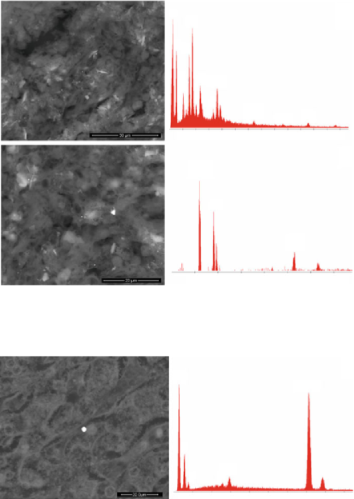
Fig. 5.1 The scanning electron microscope (SEM) image shows debris (
white spots
) of environ-
mental or occupational origin deeply embedded in a lung tissue affected by adenocarcinoma.
Among silicon-based particles (b) there are also toxic Lead-Antimony-Zinc debris. In the
y
-axis of
the EDS spectra the intensity of the signal in counts is shown and in the
x
-axis the energy in keV
a
b
C
Fe
O
CI
Fe
S
0.90 1.80 2.70 3.60
4.50 5.40 6.30 7.20 8.10
Fig. 5.2 SEM image of an Iron-Sulfur spherule detected in the liver of a patient affected by
pleural mesothelioma. The 2
m-sized Iron-Sulfur-Chlorine round-shaped debris is of combustive
μ
origin