Information Technology Reference
In-Depth Information
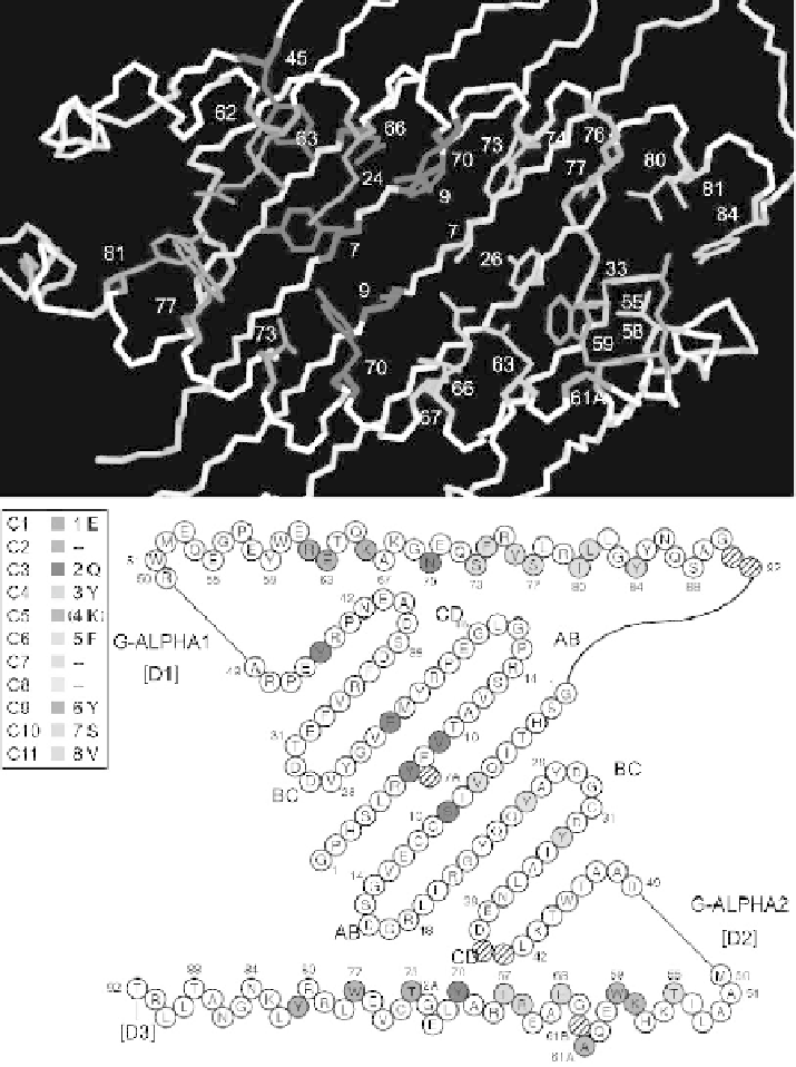
Fig. 6.
IMGT pMHC contact sites of mouse H2-K1 MHC-I and a 8-amino acid peptide (1jtr).
(A) 3D structure of the mouse H2-K1*01 groove. (B) IMGT pMHC contact sites IMGT Col-
liers de Perles. Both views are from above the cleft with G-ALPHA1 on top and G-ALPHA2
on bottom. In the box, C1 to C11 refer to contact sites (Kaas and Lefranc 2005), 1 to 8 refer to
the numbering of the peptide amino acids P1 to P8. There are no C2, C7 and C8 in MHC-I 3D
structures with 8-amino acid peptides. There is no C5 in this 3D structure as P4 does not
contact MHC amino acids (4K is shown between parentheses in the box). (A color version of
this figure appears between pages 76 and 77.)