Image Processing Reference
In-Depth Information
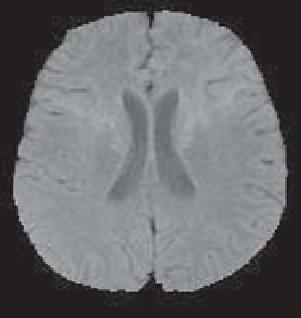
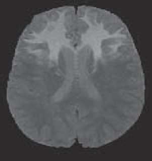
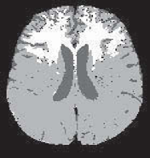
The healthy brain and the pathology cannot be distinguished in the first image and
therefore only one class can be learned, denoted by
μ
c
. In the second image, it is pos-
sible to learn two classes
μ
c
(brain) and
μ
path
(pathology). The functions
μ
c
and
μ
c
are also combined using an arithmetic mean. For the pathology,
μ
c
and
μ
path
are com-
bined using a symmetric sum defined by:
ab/
(1
b
+2
ab
). This ensures that the
pathology is not detected in the areas where
μ
path
=0and otherwise reinforces the
membership to that class, thus making it possible to include in the pathological area
the entire partial volume area where the points are comprised in part of pathological
tissue.
−
a
−
After the combination, the decision is made based on the maximum membership.
The result is illustrated in Figure 8.17.
Figure 8.17.
Two MRI acquisitions of the brain and the result of the fuzzy fusion classification
(the decision is only made locally in each point, without spatial regularization)
This example illustrates the advantage of choosing operators in an adaptive fash-
ion, based on information provided by the images about the different classes.
Search WWH ::

Custom Search