Environmental Engineering Reference
In-Depth Information
the region of cell surfaces, whereas these are absent in control cells. In living cells,
the dye molecules accumulate in the cytoplasm as granules, whereas, in starved
cells, dye molecules mainly bind on the cell surface and a very small amount is
transported to the cytoplasm.
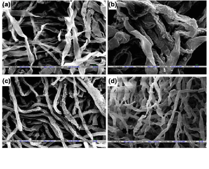
Moreover, any toxicity response, exhibited by the fungi towards the test dye,
also becomes evident through these techniques. Figure
3
shows the difference in the
mycelial structure of the fungus Aspergillus lentulus which was grown in the
presence of various dyes (Kaushik and Malik
2013
). The broad and
attened
hyphae exhibited by A. lentulus in presence ofdye Methylene Blue as compared to
that shown in the presence of Acid Navy Blue and in absence of any dye, shows the
toxicity of Methylene Blue dye to the fungi. SEM or TEM technique coupled with
Energy Dispersive X-Ray (SEM-EDX/TEM-EDX) can be a useful tool for esti-
mating and quantifying the presence of dye molecules on/inside the fungal biomass
after dye biosorption (Kaushik
2011
).
Thus, it can be concluded that the analytical techniques are an important tool to
study thedye removal process and utilization of these techniques in combination can
provide a detailed insight into the process of dye removal, its mechanism, dye
degradation pathway and study ofdegradation metabolites. This can further aid in
the development of suitable reactor design/technology for the treatment dye
ef
uents.
Fig. 3 SEM micrographs showing fungal pellets grown in different conditions a presence of
Methylene Blue (10 mg l
−
1
), b presence of Methylene Blue (50 mg l
−
1
), c presence of Acid Navy
Blue (100 mg l
−
1
) and d absence of dye (Magnication: X 5,000) (Kaushik and Malik
2013
)
Search WWH ::

Custom Search