Chemistry Reference
In-Depth Information
(a)
(b)
(c)
High
140
I
I
120
I
I
I
I
100
80
60
40
20
Low
(d)
(e)
Right
LN
Left
LN
14000
12000
Fe
3
O
4
10000
8000
6000
4000
2000
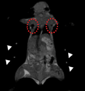
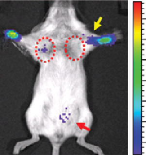
fIGure 16.31
Triple-modality imaging of radiolabelled nanoparticles: (a) optical, (b) microPET, and (c) MRI of
124
I-labelled SPIONs
injected into the front paws of a BALB/c mouse bearing a 4 T1 tumour implanted on its shoulder. Tumour: arrow; sentinel lymph node:
dotted circle; injection site: “I”; bladder: arrow; fiduciary markers: white arrow head. (d)
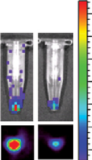
Ex vivo
luminescence (top) and microPET (bot-
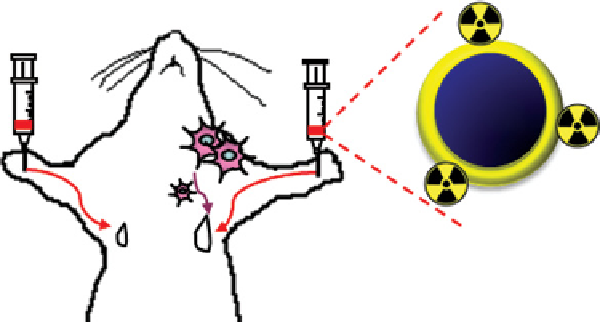
tom) images of the dissected lymph nodes. (e) Schematic diagram of tumour metastasis model and injection route of radiolabelled
nanoparticles. Reprinted with permission from Ref. [94]. (
See insert for colour representation of the figure.)
)
which dampens oscillations and reduces the power of the backscatter. However, the rigid base demonstrates a longer shelf
life and allows for covalent conjugation of targeting groups or addition of other imaging modalities and drug delivery [97].
Ultrasound and MR imaging are complementary because they combine cost-effective 2D US imaging with the 3D soft
tissue images from MR. In order to prepare MBs for use in US and MRI, SPIONs were combined with previously charac-
terised MBs [102]. First, MBs were prepared from PVA as previously reported, and SPIONs of size 8-10 nm were prepared
by a co-precipitation method. To covalently attach the SPIONs to the PVA microbubbles, amino groups were introduced on
the surface of the Fe
3
O
4
particles via silanisation with APTES. The amino groups reacted with aldehyde groups on the micro-
bubble surface, followed by reductive amination at pH 5.0 with NaBH
3
CN. Alternatively, unmodified SPIONs were added
during the PVA shell formation to physically embed them within the shell. The MBs were found to be 3.8 +/−0.6 μm in
diameter. Neither modification of the PVA MBs with iron oxide impacted the echogenicity of the MBs in a negative way
[102]. The SPIONs enabled visualisation of the microbubbles both
in vitro
and
in vivo
using MRI. Modification of MBs in
this way may be a new way of tuning the echogenicity and improving the US contrast.
16.8
MaGnetoMotIve optIcal coherence toMoGraphy (MM-oct)
Optical coherence tomography is a relatively new imaging technique that is capable of mapping 3D structures of tissue
based on optical scattering properties. The resolution is similar to that of histology. Magnetomotive optical coherence
tomography (MM-OCT) is used to image dynamic magnetic nanoprobes in biological subjects because these probes