Chemistry Reference
In-Depth Information
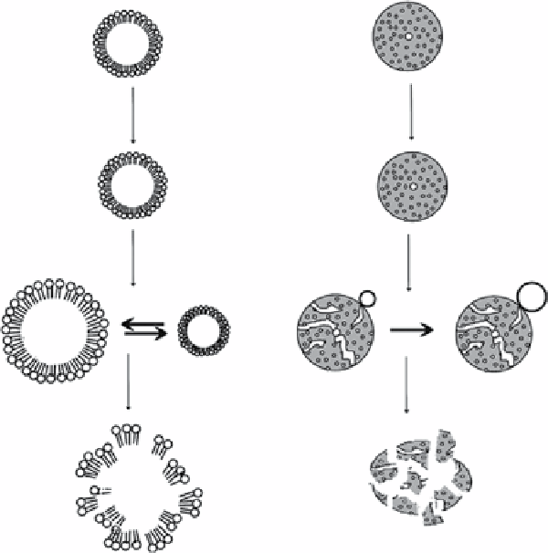
Lipid microbubble
Polymer microbubble
No US
Very-low intensity US
Low intensity US
High intensity US
fIgure 14.2
Interactions of microbubbles with US of increasing power. (Reproduced from Ref. [36], with permission from Elsevier).
vascular imaging to determine occlusion and blood flow and to detect atherosclerotic plaques. The enhancement of
microbubbles is very promising in carotid artery imaging [39], nontargeting plaque visualisation [40, 41], and accurate
stenosis assessment in carotid angiography compared to X-ray [41]. Contrast-enhanced US imaging has a unique appli-
cation in microvascular perfusion. It depicts the nonperfused regions clearly and allows the detection of perfusion insuf-
ficiency during ischemia [42], vascular occlusion [43], and tissue infarction [44] and allows the detection of vascular
insufficiency [45, 46]. other than cardiac applications, contrast-enhanced US imaging has been widely used to charac-
terise lesions in abdominal regions including liver [47-50], pancreas [51, 52], and gastrointestinal tract [53, 54]. The
in
vivo
measurement of microvascular blood flow and blood volume also allows the detection of lesions/tumours from
normal tissue [55, 56] and for the assessment of angiogenesis in tumour [57]. In addition to their vascular phase, some
microbubbles can exhibit a tissue- or organ-specific phase for improved lesion conspicuity during late enhancement
[21, 27, 29, 58-60]. Figure 14.3 illustrates the capability of identifying hepatocellular carcinoma using contrast-
enhanced US imaging, which offers a unique approach for intravascular imaging noninvasively without ionising
radiation.
14.3.2
as an orally administered us contrast agent
Diagnostic performance of abdominal US imaging is often limited by artefacts resulting from adjacent gas in the stomach
and intestines. Water, simethicone, and methylcellulose were used to displace and disperse stomach and intestinal gas for
better visualisation of upper abdomen [61]. However, it was prone to inter-subject variation and therefore results were incon-
sistent. SonoRx (bracco Diagnostics Princeton, NJ), a simethicone-coated cellulose suspension, was then developed to
improve visualisation of abdominal anatomy with reduction of gas artefacts [62, 63].
14.3.3
as a us Molecular Probe
Microbubbles can also be used in molecular imaging as a molecular probe [64]. The structure of targeted microbubbles con-
sists of a lipid/polymer shell that can become a molecular probe by incorporating different antibodies, peptides, disintegrins,
or other ligands [65, 66] (Figure 14.4).