Chemistry Reference
In-Depth Information
but not transmitted through an object/patient, as in cT imaging [4-7]. radionuclides are incorporated as part of a small metaboli-
cally active molecule to generate radiotracers such as
18
FDG, which are then intravenously injected into patients at trace dosage for
PET imaging.
18
FDG is a favourable radiotracer because it is inhibited from metabolic degradation before it decays due to the
fluorine at the 2′ position in the molecule. Upon decay, the fluorine is converted into
18
o. There is generally a short period of time
before accumulation of radiotracers into the targeted organs or tissues that are being examined, so it is important for radiotracers to
have a suitable half-life—some commonly used radionuclei have very short half-lives. Some common radionuclides used in PET
are 11-c (half-life ~20 min), 13-n (~10 min), 15-o (~2 min) and 18-F (~110 min). These are produced by a cyclotron, whereas
82-rb (76 s), which is used in clinical cardiac PET, is produced by a generator [8-9].
When a radioisotope undergoes positron emission decay (positive β-decay), it emits a positron that travels through the
tissue for a short distance (~ < 2 mm) whilst decelerating by the loss of its kinetic energy until it collides with an electron. This
results in back-to-back annihilation of
γ
-ray photons, which move in opposite directions and are emitted nearly 180 degrees
apart before being detected by scintillators and a photomultiplier tube. This type of coincidence is a true coincidence event; to
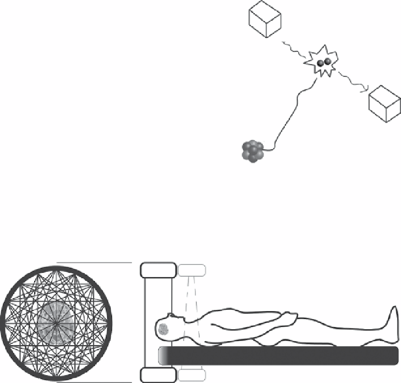
detect this, the detectors are designed like a ring that surrounds the patient during the scanning procedure. Several parallel
rings form the complete detection panel of the PET system in a cylindrical geometry (Figure 1.5).
PET has relatively high sensitivity in detecting molecular species (10
-11
- 10
-12
M), even though not all annihilation photons
are used for image reconstruction because not all coincidences are true coincidences. A coincidence event is assigned to a line
of response where the two relevant detectors are joined (detectors opposite to each other); this allows for positional information
to be located from the detected radiation without any physical collimators. This is known as electronic collimation. There are
four types of coincidence events in PET: true, scattered, random, and multiple (Figure 1.6). only true coincidence, which is
the simultaneous detection of two emissions from a single annihilation event, is useful. no other events are detected within
this coincidence time-window.
True
coincidence
γ
Detector
γ
Detector
Positron
annihilates
with electron
Two 511 photons are
emitted simultaneously
in opposite direction
Pet scanner
Typical conguration:
Emission
positron
Whole-body (patient port around 60 cm and FOV around 15 cm)
Scintillator crystals coupled to photomultiplier
Unstable nucleus
Cylindrical geometry
Other congurations for
special-purpose applications:
24-32 rings of detector crystals
Hundreds of crystal/ring
Brain imaging
Animal PET
LORS
PET
CT
Mammography
Other
True
FIgure 1.5
Typical configuration of a PET scanner.