Biomedical Engineering Reference
In-Depth Information
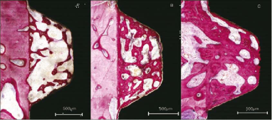
Figure 9.4.
Histologic view of enhanced bone healing adjacent to a chemically modified implant surface with bone formation
(A) after 2 weeks of healing, (B) after 4 weeks of healing, and (C) after 8 weeks of healing. Reproduced from Buser et al. [10] , with
permission of the International and American Associations for Dental Research.
One of the other materials being tested
around implants is BMP. In one study, rhBMP-
2
, either as part of a collagen sponge or mixed
with a polylactide-glycolide polymer, was
placed around dental implants inserted in the
partially edentulous mandible of dogs [
].
Half of the defects were covered with a nonre-
sorbable membrane. In control sites only carrier
was used. Sites that contained BMP had signifi -
cantly more bone in the defects and against the
implant surface than control sites. Early in the
study, the membrane-covered sites had less new
bone than the uncovered sites. At later times,
the membrane-covered sites had more bone.
Furthermore, sites with collagen had more bone
than sites with the synthetic carrier.
19
,
20
9.4 Bone Regeneration in
Areas Insufficient for
Implant Placement
As implant therapy develops, more implants
are inserted into sites that lack suffi cient bone
to support an implant. It therefore has become
necessary to regenerate lost alveolar bone tissue
with the aid of guided bone regeneration
(GBR).
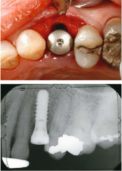
Figure 9.5.
Clinical view (top) and radiographic view (bottom)
of the placement of a dental implant into a tooth extraction
socket. The clinical view shows the residual space between the
implant and alveolar bone requiring bone grafting.