Information Technology Reference
In-Depth Information
The
I
b
IN population activity with dopamine was given by
dX
i
dt
=
φ
A
11
(
15
−
X
i
)
F
i
−
X
i
DA
11
(
0
.
8
+
2
.
2max
(
X
j
,
0
))
,
(11.9)
where
F
i
is the feedback activity of force-sensitive Golgi tendon organs.
11.7 Simulated Effects of Dopamine Depletion on the Cortical
Neural Activities
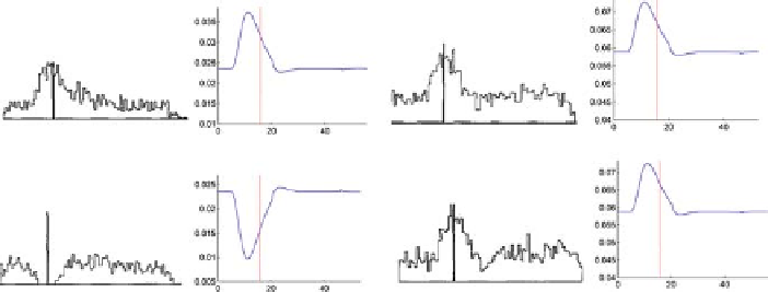
Figures 11.5 and 11.6 show qualitative comparisons of experimental and simu-
lated neuronal discharges of reciprocal and bidirectional neurons in normal and
dopamine-depleted conditions, respectively. It is clearly evident an overall reduction
of firing intensity [23, 36], a reduced rate of change of neuronal discharge [23, 36],
a disorganization of neuronal activity (neuronal direction specificity is markedly
reduced) [23], and an increase in baseline activity (in the normal case the base-
line activity was 0.05, whereas in dopamine depleted the baseline activity increased
to 0.07) [23]. Figure 11.8 shows a qualitative comparison of abnormal cellular re-
sponses of GPi neurons to striatal stimulation in MPTP-treated monkeys (column
1 of Fig. 11.8) and simulated GPi neuronal responses (column 2 of Figure 11.8).
A
C
B
D
Fig. 11.6: Comparison of peristimulus time histograms (PSTH) of reciprocally or-
ganized neurons (column 1; reproduced with permission from [23, Fig. 4A, p. 182],
Copyright Springer-Verlag) in area 4, simulated area's 4 reciprocally organized
phasic (DVV) cell activities (column 2), PSTH of area's 4 bidirectional neurons
(column 3; reproduced with permission from [23], Fig. 4A, p. 182, Copyright
Springer-Verlag) and simulated area's 4 co-contractive (P) cells activities (column
4) for a flexion (
A
and
C
) and extension (
B
and
D
) movements in MPTP-treated
monkey. The
vertical bars
indicate the onset of movement. Note that the triphasic
pattern is disrupted: Peak AG1 and AG2 bursts have decreased, and ANT pause is
shortened.