Information Technology Reference
In-Depth Information
CRT
T
A
T
B
NEURONAL ACTIVITY
EMG ACTIVITY
PMT EMT
DISPLACEMENT
VELOCITY
RT
MT
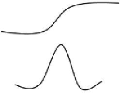
Fig. 11.3: Schematic representation of the neuronal, electromyographic, and kine-
matic variables. CRT: cellular reaction time; PMT: premotor time; EMT: electrome-
chanical time; RT: reaction time; MT: movement times;
T
A
: time of neuronal dis-
charge prior to movement onset;
T
B
: duration of neuronal discharge after movement
onset.
arm displacement and the duration of the neuronal activity from the onset of move-
ment and the time where the level of activity returned to resting levels (see Fig. 11.3
for a schematic description) were increased. Similarly, Doudet and colleagues [23]
reported that the mean duration of neuronal discharge in area 4 preceding the onset
of movement was slightly affected in the MPTP-treated animals, whereas the mean
duration of neuronal discharge following the onset of movement was significantly
increased.
11.5.4 Prolongation of Behavioral Simple Reaction Time
Benazzouz et al. [2] trained monkeys to perform a rapid elbow movement (
40) of
extension or flexion in response to an auditory signal. EMG activity was recorded
with intramuscular electrodes 500 ms before and 1,500 ms after the beginning of
the auditory signal, before and after an MPTP lesion of the substantia nigra pars
compacta (SNc). They reported that the behavioral simple reaction time (cellular
reaction time
>
mean duration of neuronal discharge before movement onset (TA;
see Fig. 11.3) after a nigral MPTP lesion was significantly increased for both exten-
sion and flexion movements. Similarly, Doudet et al. [24, 23] and Gross et al. [36]
observed a significant change in the mean values of the simple reaction time (RT)
+


