Biomedical Engineering Reference
In-Depth Information
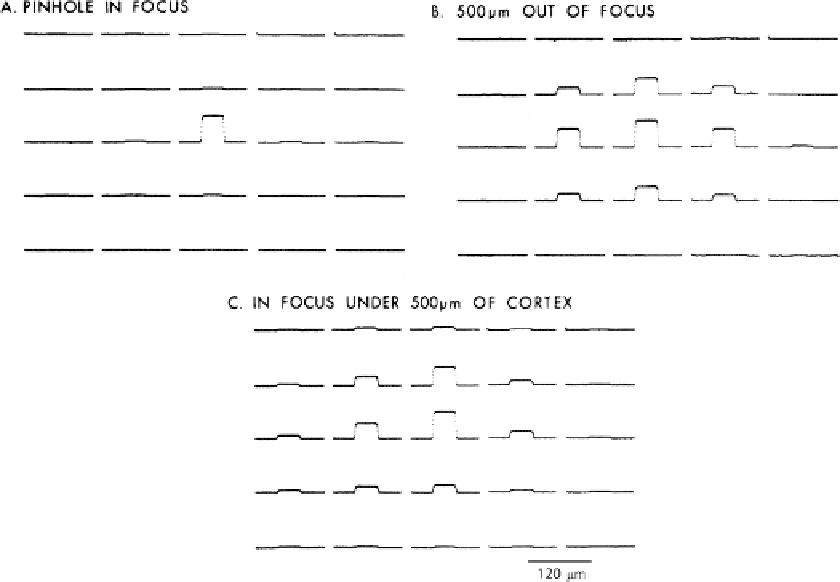
Fig. 3.12. Effects of focus and scattering on the distribution of light from a point source onto a camera. The data were
acquired using a 464 element photodiode array. (
A
)A40μm pinhole in aluminum foil covered with saline was illuminated
with light at 750 nm. The pinhole was in focus. More than 90% of the light fell on one detector. (
B
) The stage was moved
downward by 500 μm. Light from the out-of-focus pinhole was now seen on several detectors. (
C
) The pinhole was in
focus but covered by a 500 μm slice of salamander cortex. Again the light from the pinhole was spread over several
detectors. A 10 × 0.4 NA objective was used. Kohler illumination was used before the pinhole was placed in the object
plane. The recording gains were adjusted so the largest signal in each of the three trials would be approximately the
same size in the figure. Redrawn from Orbach and Cohen
(61)
.
images from intact vertebrate preparations with much better spa-
tial resolution than is achieved with wide-field microscopy. The
two-photon microscope has been successfully used to monitor
changes in calcium concentration inside small processes of neu-
rons
(78)
and, in many individual cells, after bulk loading the
AM ester calcium dyes (
(38)
; Example 2 above). Using a scanned
line confocal microscope from Prairie Technologies (Middletown,
WI), Jeff Magee and collaborators (personal communication)
were able to measure voltage-sensitive dye signals from hippocam-
pal cell dendrites at a frame rate of 3 kfps. Slower voltage-sensitive
dye signals were measured confocally much earlier
(79)
.
Because the signal-to-noise ratio in a shot noise limited mea-
surement is proportional to the square root of the number of
photons converted into photoelectrons, quantum efficiency is
important. Silicon photodiodes and CCD and CMOS cameras
can have quantum efficiencies approaching the ideal (1.0) at
4.4. Cameras
4.4.1. Quantum Efficiency