Biomedical Engineering Reference
In-Depth Information
EPSP-AP pairing activity pattern that induces persistent changes
in synaptic efficacy
(12)
.
2.2. Activity of Many
Individual Neurons in
the Mouse Primary
Somatosen-
sory/Visual Cortex in
Response to Sensory
Stimulation (Fig. 3.3,
Middle Panel)
Nervous systems are made up of large numbers of neurons and,
many of these are active during the generation of behaviors. The
original motivation for developing optical methods for monitor-
ing activity was the hope that they could be used to record activity
of many neurons simultaneously during behaviors
(33)
. Obtain-
ing information about the activity of many cells is essential for
understanding the roles of the individual neurons in generating
behavior and for understanding how nervous systems are orga-
nized.
One simple and widely used method to monitor neuronal
activity relies on imaging the neuron's intracellular Ca
2
+
con-
centration. Indeed, in living cells, most depolarizing electrical
signals are associated with Ca
2
+
influx caused by the activation
of different types of voltage-gated Ca
2
+
channels, abundantly
expressed in the nervous system
(34, 35)
. Such signals are often
further amplified by Ca
2
+
release from intracellular Ca
2
+
stores
(36)
. The easiest technique to monitor activity of many individ-
ual neurons by imaging their intracellular Ca
2
+
concentration uses
a membrane permeant acetoxymethyl (AM) ester form of a Ca
2
+
indicator dye. Such dyes were first introduced by R.Y. Tsien
(37)
and widely used ever since. For the vertebrate brain in vivo, the
method allowing imaging of neural ensembles with single cell res-
olution was introduced by Stosiek et al.
(38)
. The technique was
originally developed for imaging layer 2/3 neurons in the mouse
cortex (
Fig. 3.6
), and was successfully applied later to stain dif-
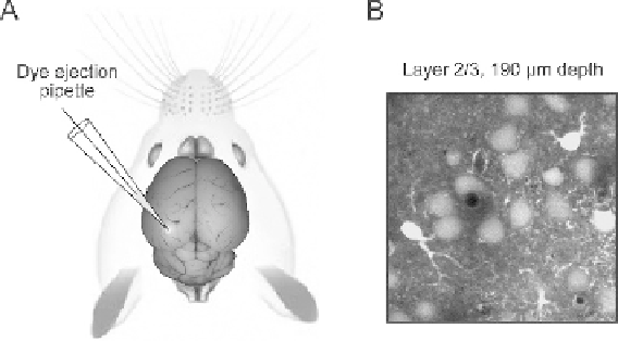
Fig. 3.6. A, a schematic drawing illustrating in vivo multi cell bolus loading of cortical
neurons with a calcium indicator dye. The membrane-permeant dye is pressure-injected
from the patch pipette into the extracellular space. Subsequently, it diffuses into the cells
where it is deesterified by intracellular esterases. B, an example of image quality. The
cells in layer 2/3 of the primary visual cortex were stained using multi cell bolus loading
and visualized using two-photon imaging.