Biomedical Engineering Reference
In-Depth Information
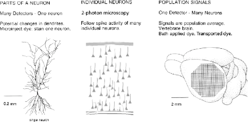
Fig. 3.3. Schematic drawings of the three kinds of measurements described as examples. Left, an individual cortical
hippocampal CA1 pyramidal cell. Each pixel of an
80
80
pixel CCD camera would receive light from a small part of
the dendrite, axon, or cell body of the neuron. An optical measurement of membrane potential would provide important
information about how the neuron converts its synaptic input into its spike output. Middle, a drawing of cells in a
vertebrate cortex. 2-photon imaging allows imaging of the fluorescence of many individual neurons stained with a
calcium sensitive dye. Right, a vertebrate brain with a superimposed pixel array. Each pixel of the array would receive
light from thousands of cells and processes. The signal would be the population average of the change in membrane
potential in those cells and processes.
×
activation of many neurons in wide-spread brain areas; optical
imaging allow simultaneous measurement of population signals
from many areas. In these three instances, optical recordings have
provided kinds of information about the function of the nervous
system that were previously unobtainable.
2. Three
Examples
Understanding the biophysical properties of single neurons and
how they process information is fundamental to understanding
how the brain works. At present, however, the detailed func-
tional structure of nerve cells is not fully understood. Part of
the explanation for this incomplete understanding is that neurons
are exceedingly complex. It is widely recognized that dendritic
membranes of many vertebrate CNS neurons contain active con-
ductances such as voltage-activated Na
+
,Ca
2
+
and K
+
channels
(e.g.
(15-20)
). An important consequence of active dendrites is
that regional electrical properties of branching neuronal processes
will be extraordinarily complex, dynamic, and, in the general case,
impossible to predict in the absence of detailed measurements. To
obtain such a measurement, one would, ideally, like to be able
2.1. Processes of an
Individual Neuron
(Fig. 3.3, Left Panel)