Biomedical Engineering Reference
In-Depth Information
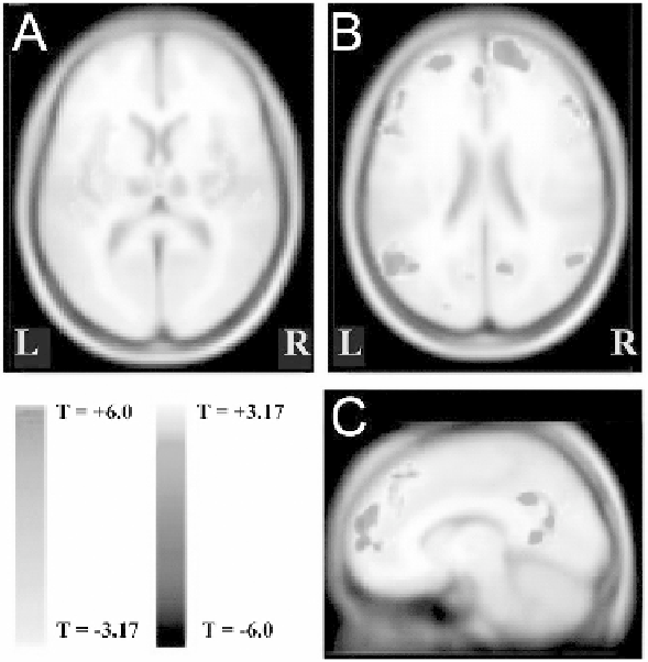
Fig. 9.3. fMRI changes in group analysis of generalized spike-wave seizures (15
patients with idiopathic generalized epilepsy). (
A
) Significant increases are shown in the
bilateral thalami and in several cortical areas. (
B, C
). Decreases are shown in bilateral
interhemispheric regions, lateral frontal cortex, and parietal association cortex. Modified
with permission from Gotman et al, 2005 PNAS
(69)
. Color version of this figure can be
viewed in the original publication
(69)
at http://www.pnas.org/.
data with the behavioral deficits often associated with absence
seizures, simultaneous behavioral testing may allow researchers to
understand the relationship between fMRI changes and behav-
ioral changes
(28, 68)
. Thus, it will be critical in further studies to
directly link fMRI signal changes with behavioral deficits. Finally,
techniques to date have not necessarily differentiated BOLD
activity based on seizure duration. CBF and brain metabolism
may differ in brief SWD as opposed to in prolonged seizures
or absence status epilepticus. Furthermore, fMRI signal changes
and the spatial heterogeneity of SWD amplitude in different brain
regions based on EEG (or MEG) have not yet been correlated.
6. Animal fMRI
Studies of
Spike-Wave
Animal studies are vital companions to human studies and pro-
vide advantages for studying epilepsy and conducting fMRI
experiments. Animal models of epilepsy often allow investigators