Biomedical Engineering Reference
In-Depth Information
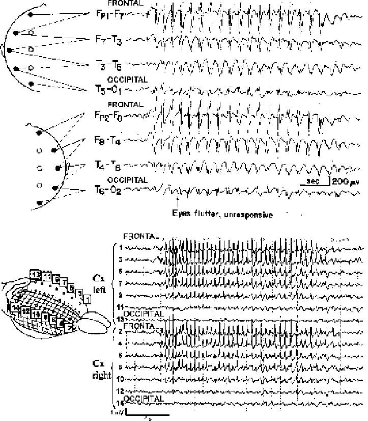
Fig. 9.1. Typical spike-wave EEG discharges during absence seizures show some regions intensely involved, and others
relatively spared by seizures. (
A
) EEG recordings of a typical absence seizure from a 7-year old girl, reveals bilateral
synchronous 3-4 Hz spike-wave discharges, with an anterior to posterior amplitude gradient. (Inset of electrode positions
modified with permission from Fisch, B.J. (1991) Spehlmann's EEG Primer. Elsevier, Amsterdam. EEG recording modified
with permission from Daly, D.D. and Pedley, T.A. (1990) Current Practice of Clinical Electroencephalography. Raven Press,
New York.). (
B
) Electrocorticography from the surface of the WAG/Rij rat cortex during spike-and-wave seizures exhibits
intense involvement of the anterior cortex and relative sparing of the occipital lobes. (Reprinted with permission from
Meeren et al., 2002. Copyright 2002, Society for Neuroscience.)
Electroencephalogram (EEG) recordings during typical
absence seizures reveal large-amplitude, bilateral 3-4 Hz
spike-wave discharges (
Fig. 9.1
). SWDs are usually bilaterally
symmetric, however, left or right amplitude preponderance occurs
occasionally
(7)
. Although absence seizures are considered gen-
eralized epileptic events, SWD amplitude in humans is seen