Biomedical Engineering Reference
In-Depth Information
pre-synaptic
neuron
pre-synaptic
potentials
capillary
Na
+
glucose
&
oxygen
Na
+
ATP
K
+
glucose
&
oxygen
Ca
2+
synaptic
field
potentials
ATP
K
+
Na
+
Na
+
Na
+
Na
+
astrocyte
K
+
ATP
Ca
2+
Ca
2+
post-synaptic
potentials
Na
+
nitric oxide
and others
post-synaptic
neuron
capillary
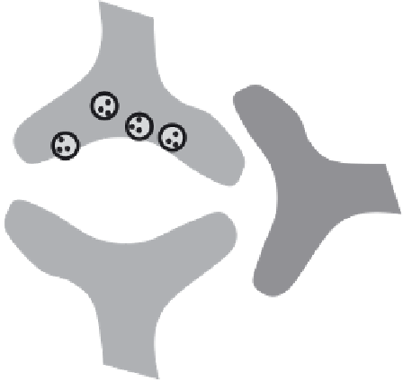
Fig. 1.1. Cytological association between microvasculature with neurons and astrocytes in the glutamatergic synapse.
metabolic, and/or hemodynamic changes, it is important to clas-
sify the underlying basic processes with appropriate spatial and
temporal scales.
Functional integrity of the working brain is maintained
by electrical communication amongst an enormous number of
neurons with active partnership provided by astrocytes
(4, 5)
.
Cytological association of neurons and astrocytes with the
microvasculature (
Fig. 1.1
) provides the framework that links
activities at the nerve terminal to energy demand
(6, 7)
and blood
flow
(8, 9)
. In the mammalian cerebral cortex, glutamate is the
major excitatory neurotransmitter, whereas
-amino butyric acid
(GABA) is its conjugate inhibitory partner. Together, they consti-
tute nearly 90% of cortical neurons
(10)
. Glutamate metabolism
plays a central role in both glutamatergic and GABAergic synapses
because glutamate is a precursor of GABA and it is a constituent
of both neurons and astrocytes
(11, 12)
. Thus, featuring proper-
ties of the glutamatergic system seem to be an appropriate starting
point for this discourse.
γ
2. Activities at the
Nerve Terminal
Electrical communication between cells at the glutamatergic
nerve terminal is characterized by 1-2 ms epochs of cellular
discharge (or depolarization) which are followed by quiescent