Biomedical Engineering Reference
In-Depth Information
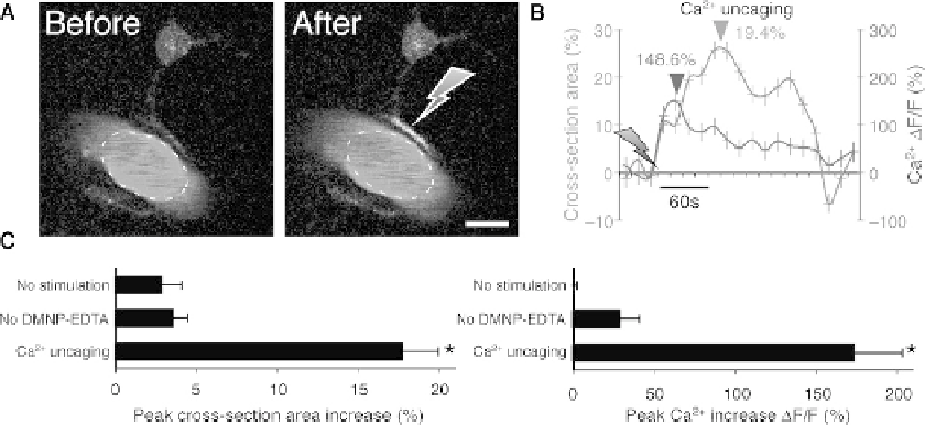
Fig. 5.4. Vasodilation triggered by uncaging (
A
) Upper panel: Two-photon images of a vascular astrocyte, which dis-
played Ca
2
+
increases in the endfoot following photolysis of caged Ca2+. Astrocytes were loaded with the Ca
2
+
indica-
tor dye, rhod-2/am and DMNP-EDTA/am, whereas the vasculature was stained with FITC-dextran. Gray arrow indicates
the position of photostimulation. Scale bar, 10 μm. (
B
) Time-course tracings show that photostimulation caused a rapid
Ca
2
+
increase and arterial vasodilation. Arrows indicate time of photolysis stimulation. Dark arrowhead indicates the
peak of Ca
2
+
elevation, and light arrowhead indicates peak dilation. (
C
) Summary histograms of maximum arterial
cross-section area (left panel) and maximum Ca
2
+
increase (right panel) without photostimulation, with photostimula-
tion but omitting DMNP-EDTA/am loading, and with DMNP-EDTA loading and photolysis. Maximum increase within 1 min
after photostimulation was measured relative to baseline before stimulation. Mean ± s.e.m. * P
<
0.01 compared to
no-stimulation, Tukey-Kramer.
endfeet, indicated that the primary mechanism by which astro-
cytes mediated vessel dilation is through release of PGE2 in a
COX-1-dependent pathway
(51)
(
Fig. 5.4
).
Interestingly, several lines of work have documented that
the changes in blood flow and BOLD signal detected during
brain activation have a stronger correlation with synaptic inputs
compared with spiking activity
(52, 53)
. Indeed, local spiking
activity does not appear to play a central role in functional hyper-
emia
(53)
. Thus, activity-dependent vascular signals reflect exci-
tatory synaptic activity or synaptic release of glutamate
(54, 55)
.
Similarly, astrocytes respond to the synaptic inputs, or glutamate
released from synaptic terminal during sensory stimulation, and
local spiking activity has no effect upon astrocytic Ca
2
+
eleva-
tion
(9)
. Thus, astrocytic Ca
2
+
signaling may represent a signal-
ing pathway that locally integrates synaptic inputs and controls
the microvasculature. Astrocytes detect presynaptic neurotrans-
mitters release and display increases in intracellular Ca
2
+
. In turn,
astrocytic Ca
2
+
signaling activates PLA2 resulting in release of
vasoactive COX-1 metabolic products. Other pathways, such as
NO, adenosine and pH may in parallel contribute to functional
hyperemia. Several of these pathways may be similar to gluta-
matergic pathways which require astrocytes as a necessary inter-
mediary linking synaptic activity to vasodilatation.