Biomedical Engineering Reference
In-Depth Information
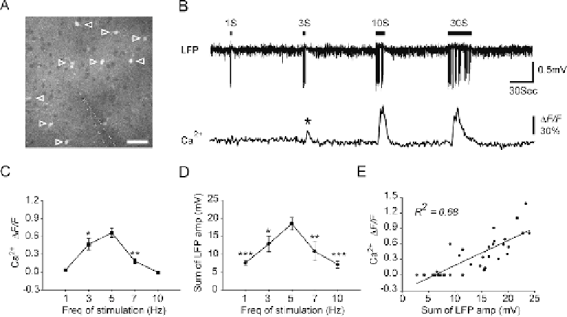
Fig. 5.3. Whisker stimulation evokes astrocytic Ca
2
+
increases in mouse barrel cortex. (
A
) Two-photon fluorescence
image taken from layer 2/3 of mouse barrel cortex (∼150 μm below the pial surface), showing that astrocytes were
labeled with Ca
2
+
indicator fluo-4 AM (open arrowheads). Neuronal cell bodies appear as dark, round area, due to their
lack of uptake of fluo-4 AM. The location of recording electrode is indicated by white dashed line. Scale bar, 30 μm.
(
B
) 5 Hz whisker stimulation induced astrocytic Ca
2
+
increases as a function of the stimulation duration. Upper trace,
LFP recorded from recording electrode shown in panel A. Lower trace, averaged relative fluorescent change
F/F in
the astrocytic cell bodies indicated in panel A. (
C
) Mean increase in fluo-4 emission after 9 s of different frequencies
of stimulation. (
D
) Summed LFP amplitude within the first 9 s of whisker stimulation as a function of the frequency of
stimulation. Data are presented as mean ± s.e.m. * P
<
0.05, ** P
<
0.01 and *** P
<
0.001 compared with 5 Hz whisker
stimulation induced responses. One-way analysis of variance with Dunnett's test. (
E
) Correlation between the summed
LFP amplitude within the first 9 s and the mean increase in fluo-4 emission 9 s after the onset of stimulation (R
2
=0.68,
P
<
0.001).
changes of somatic fluorescent signal reached 3 standard deviation
of baseline fluorescent activity. Notably, 1-s stimulation failed to
evoke astrocytic Ca
2
+
signaling, whereas 3-s stimulation was suf-
ficient to induce astrocytic Ca
2
+
elevation (asterisk) (
Fig. 5.3B
).
In addition, the amplitude of astrocytic Ca
2
+
increases was
similar in response to 10-s and 30-s stimulation, while the dura-
tion of astrocytic Ca
2
+
elevation was longer during 30-s stim-
ulation. This evidence indicates that the amplitude of astrocytic
Ca
2
+
elevation was mainly determined by the neuronal activity
during the first
∼
10 s of stimulation (
Fig. 5.3B
). Most astrocytes
showed a single increase in Ca
2
+
in response to 1 min of whisker
stimulation (72 cells from 13 mice), whereas others showed a sec-
ond Ca
2
+
increase (11 cells from 13 mice).
In addition to the effects of the duration of stimulation on
astrocytic Ca
2
+
elevations, astrocytic Ca
2
+
responses are also a
function of the frequency of stimulation. During 1 Hz whisker
stimulation, only 3 of 37 astrocytes tested in 9 mice showed an
increase in Ca
2
+
(9)
. Rapid sensory adaptation, or a decrease