Geology Reference
In-Depth Information
Stable isotope data
dissolved carbonate ions derived from organic
matter as a result of microbial activity.
Using the
The carbon and oxygen isotopic compositions of
the bulk sediment remain basically unchanged
until 18 cm depth (Fig. 3c; Table 1). As the
percentage of dolomite increases to 100% at
20-23 cm depth, the
18
O palaeotemperature equation
calibrated for dolomite (Vasconcelos
et al
., 2005),
we can calculate the diagenetic formation temper-
ature of the dolomite from 20 to 23 cm depth. With
an average
18
O values increase slightly
as a new isotopic equilibrium with the pore-
water is obtained, whereas the
18
O V-SMOW of 2.31‰ for the pore-
18
O V-PDB of 3.14‰ for the dolomite,
the calculated temperature for the early diagen-
esis is 21°C, which corresponds well to an average
value observed with fi eld measurements at this
depth below the sediment-water interface.
water and
13
C values
become relatively much more negative over the
same interval. The large change in
13
C indic-
ates isotopic re-equilibration with the input of
HMC
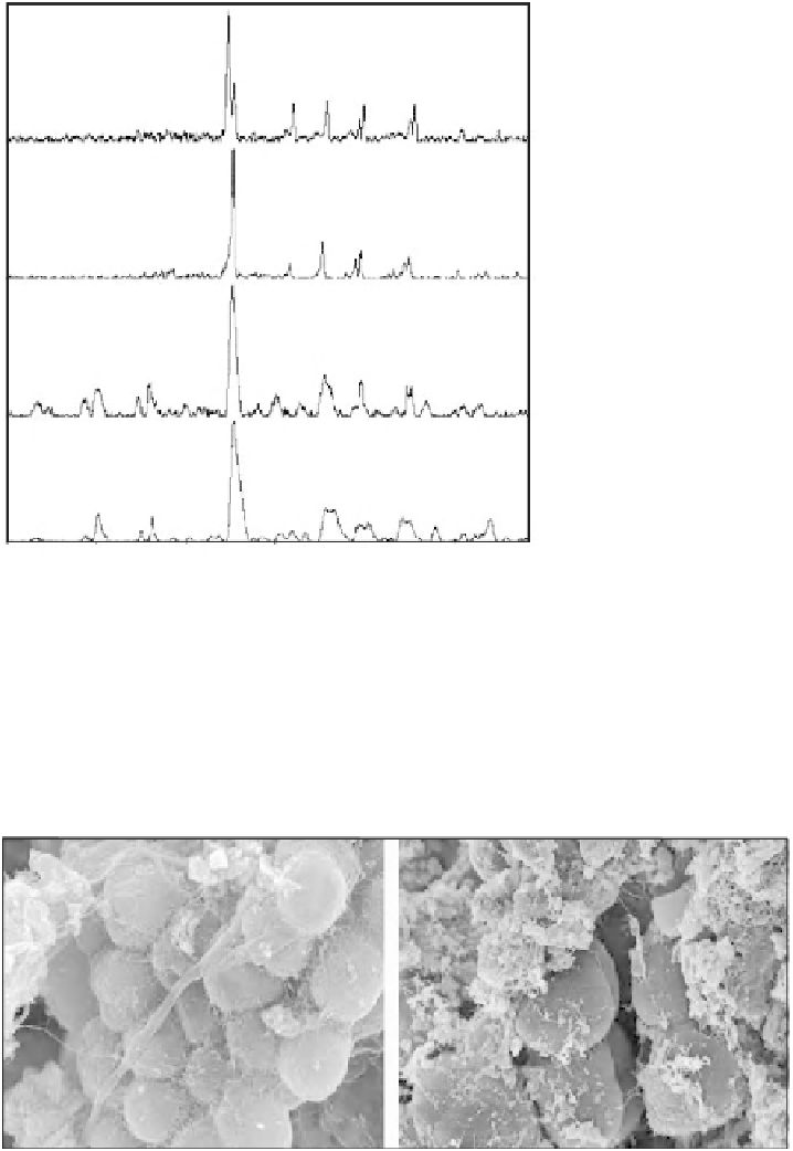
(a)
Scanning electron microscopy of sediment
With SEM imaging it is possible to detect
microbial colonies associated with the sedimen-
tary carbonates in Brejo do Espinho. Figure 5
shows detailed views of the sediment sampled at
5 cm depth, which indicate the presence of cells
within a matrix of nanocrystals (Fig. 5a) and coc-
coid colonies shown typically embedded in EPS
(Fig. 5b). Figure 6 is an SEM photomicrograph of
the well-crystallized rosettes comprising nano-
crystals found in the 100% dolomite sediment
from 21 cm depth. These SEM images support
the hypothesis that microbes are associated with
primary dolomite precipitation in the sediment
near the sediment-water interface and the
re-equilibration of the crystals with early diagen-
esis at shallow depths. This ageing process was
also observed in the nearby Lagoa Vermelha
sediment, where it occurs under reducing
conditions in the presence of sulphate-reducing
bacteria (Vasconcelos & McKenzie, 1997).
D
(b)
D
(c)
D
HM
(d)
D
HM
5
15
25
35
45
55
65
θ
)
Diffraction angle (º2
Fig. 4.
X-ray diffractograms of Brejo do Espinho samples
from (a) 5 cm depth showing a mixture of high-Mg calcite
and dolomite and (b) 21 cm depth showing 100% dolo-
mite. X-ray diffractograms of biogenic crystals formed
by (c)
V. marismortui
and (d)
Marinobacter
sp. (Note:
D = dolomite, HMC = high-magnesium calcite and
HM = hydromagnesite.)
Aerobic culture experiments
Laboratory culture experiments were performed
using the two heterotrophic microorganisms,
V. marismortui
and
Marinobacter
sp
.
(strains
(a)
(b)
Fig. 5.
SEM photomicrographs
of Brejo do Espinho sediment
taken from 5 cm in short core.
(a) Coccoid bacterial cells embed-
ded in extracellular organic
matter. (b) Close-up of the fi ne-
grained sediment showing the
presence of cells (marked with
arrows).
1
μ
m
1
μ
m