Agriculture Reference
In-Depth Information
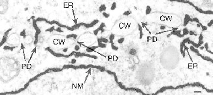
Figure 5.3
Cytokinesis in the root tip of
Zea mays
showing formation of primary plasmodesmata by
endoplasmic reticulum entrapment in the developing cell plate. The tissue has been impregnated with
zinc iodide/osmium tetroxide; this technique deposits osmium on the cisternal space of
membrane-bound organelles such as the endoplasmic reticulum. Smooth domains of the endoplasmic
reticulum crossing the growing cell plate indicate areas where primary plasmodesmata will form,
constricted endoplasmic reticulum tubules (desmotubule) can also be seen between areas of the
developing cell wall. ER, endoplasmic reticulum; CW, cell wall; PD, plasmodesmata; NM, nuclear
membrane. Image courtesy of C. Hawes, Oxford Brookes University. Bar: 25 nm.
each developing phragmoplast, allowing cytoplasmic and endomembrane continu-
ity to occur between daughter cells (see Fig. 5.3; Mezitt & Lucas, 1996).
Secondary
plasmodesmata
are defined as those that form
de novo
across existing cell walls
(Lucas
et al.
, 1993; Ehlers & Kollmann, 2001). Hepler (1982) provided the first
detailed electron microscopy study of primary plasmodesmata formation during
cell-wall deposition in roots of
Latuca sativa
.Atthe final stage of cytokinesis, a
cell wall is formed to partition the two daughter cells. It is generally accepted that
primary plasmodesmata are formed at sites where endoplasmic reticulum tubules
become trapped within the fusing Golgi-derived vesicles of cell wall material form-
ing the cell plate (Porter & Machado, 1960; Hepler, 1982; Staehelin & Hepler, 1996).
The cytoplasmic strands become constricted during cell plate growth, and the en-
doplasmic reticulum tubules become transformed into the desmotubule, retaining
endomembrane continuity between the two daughter cells (Ehlers & Kollmann,
2001). Overall (1999) has suggested that the dynamin-like molecule phragmoplas-
tin, which has been identified in forming the cell plate (Gu & Verma, 1996), may
induce the production of the tightly curled endoplasmic reticulum membrane tube
as it passes through the plasmodesma.
Although plasmodesmata formed during cytokinesis are randomly distributed,
they are typically grouped into pit fields in the fully elongated cell (Fisher, 2000). The
number of primary plasmodesmata laid down in a given wall has been found to accu-
rately predict the subsequent cell wall expansion that will take place (Overall, 1999).
Little is known about how this process is regulated, although short-term treatment