Biomedical Engineering Reference
In-Depth Information
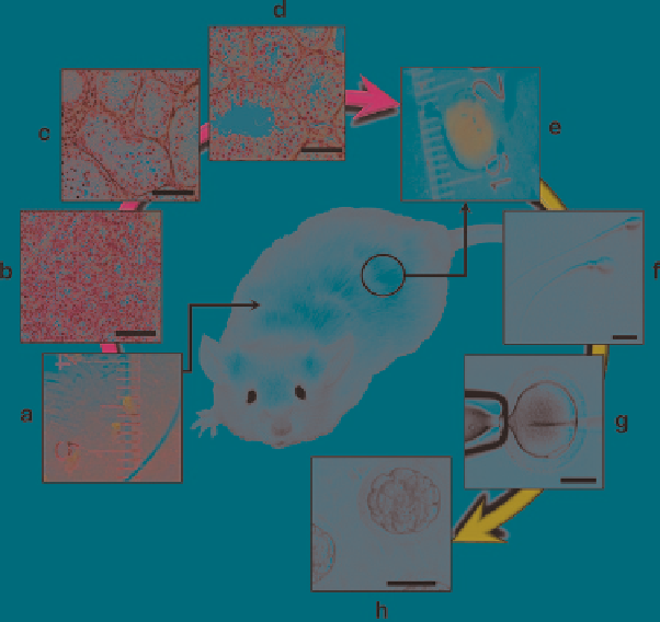
Fig. 10.1
Ectopic xenografting of immature primate testis tissue into immunodeficient mice.
Fragments of immature donor testis (~1 mm
3
) transplanted under the dorsal skin of immunodefi-
cient mice (
a, b
) are able to survive and respond to gonadotropins. As a result, testis tissue under-
goes complete development, including formation of fertilization competent sperm (
c-f
). Once
testis xenografts are collected (
e
) they can be used for analysis or to obtain sperm for ICSI (
g
) and
embryo production (
h
).
bars
equal 50 mm (
b-d, g, h
) or 10 mm (
f
)
Since the time of sperm retrieval is often many years earlier than their estimated
use, excellent protocols for cryopreservation and cryostorage are important prereq-
uisites when testicular grafting will become a clinical tool. To maintain several
options for future use of the cryopreserved material, testicular tissue is best cryo-
preserved as small fragments as well as enzymatically dispersed single cell suspen-
sions. This will maintain options to use the intact tissue as grafts or to create a
single cell suspension for reaggregation of testicular tissue or isolation of sper-
matogonial stem cells for
in vitro
approaches or germ cell transplantation. Protocols
for cryopreservation of cell suspensions and testicular fragments from adult and
cryptorchid testes using propanediol, glycerol, ethylene glycol, or DMSO were
described (Brook et al.
2001
; Keros et al.
2005, 2007
; Kvist et al.
2006
). However,
none of these studies had assessed the
in vivo
stem cell capacity of the cryopre-
served and thawed primate spermatogonia. Xenografting was applied to optimize
procedures for cryopreservation of testicular tissue (Jahnukainen et al.
2007a
). In
addition to showing that immature primate testis tissue can best be cryopreserved
Search WWH ::

Custom Search