Biomedical Engineering Reference
In-Depth Information
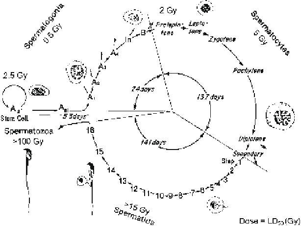
Fig. 9.2
Sequence, kinetics, and radiation sensitivities of spermatogenic cells in the mouse. The
LD
50
is the radiation dose necessary to kill 50% of the cells [modified with permission from
(Meistrich et al.
1978
)]
susceptible to cytotoxic agents (Fig.
9.2
) (Oakberg
1957
). The later stage germ cells
(spermatocytes and especially spermatids) are less sensitive to killing by most of
these cytotoxic agents (Meistrich et al.
1982
; Oakberg and Diminno
1960
). The
somatic cells of the testis also survive most cytotoxic therapies; however, these cells
may suffer functional damage (Zhang et al.
2006
).
After cytotoxic treatment, the time course of changes in sperm count depends on
the sensitivities of the different spermatogenic cells, and their kinetics and effi-
ciency of maturation to sperm in the testicular environment. Once the progeny of
the stem spermatogonia differentiate to the point at which cells are related to spe-
cific stages of the cycle of the seminiferous epithelium (A
1
spermatogonial stage in
rodents, B spermatogonial stage in primates), they progress with the same kinetics
as in the normal testis.
Because of the relative resistance of the later stage germ cells, the immediate
effect of cytotoxic exposure on sperm count is minor (at low doses) or gradual
(at higher doses) (Fig.
9.3
). However, at the times that the differentiating
spermatogonia would have become sperm, ranging from 35 days in mice to 60 days
in humans, sperm counts often decline dramatically. This occurs with the doses of
a highly gonadotoxic agent like radiation shown in Fig.
9.3
. Even mildly gonado-
toxic forms of chemotherapy, which do not affect stem cells or the recovery of
spermatogenesis from the stem cells, can cause transient reductions in sperm count
lasting until 2-3 months from the end of treatment because they kill differentiating
spermatogonia (Lu and Meistrich
1979
; Meistrich et al.
1997a
).
Search WWH ::

Custom Search