Biomedical Engineering Reference
In-Depth Information
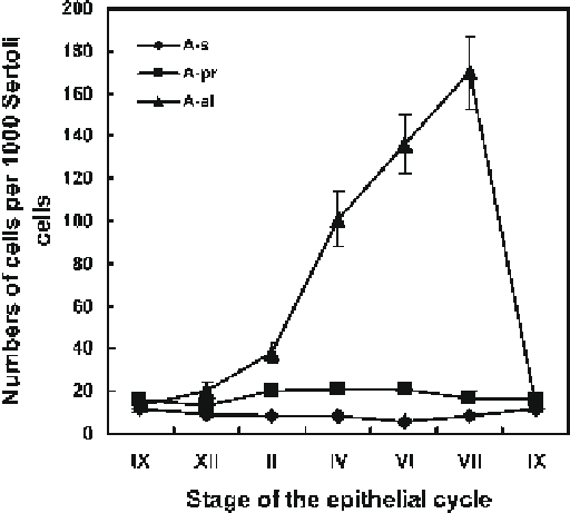
Fig. 4.4
Numbers of A
s
, A
pr
, and A
al
spermatogonia throughout the stages of the cycle of the
seminiferous epithelium in the Chinese hamster. Data are from Lok et al. (
1982
)
26 in 101xC3H mice to 60 per 1,000 Sertoli cells in C3H mice (Tegelenbosch and
de Rooij
1993
; Huckins and Oakberg
1978
).
4.6
Cell Cycle Characteristics of A
s,p r, al
Spermatogonia
As first shown by Huckins and Kopriwa, it is possible to carry out autoradiography
on whole mounts of seminiferous tubules (Huckins and Kopriwa
1969
). Using this
technique after
3
H-thymidine administration, cell cycle times have been established
of all types of spermatogonia in the rat (Huckins
1971a, d
) and the Chinese hamster
(Lok and de Rooij
1983a
; Lok et al.
1983
). Again advantage has been taken of the
wave of spermatogenesis and the synchronous behavior of the A1-B spermatogonia.
Shortly after injection of the
3
H-thymidine all A1-B spermatogonia in S-phase incor-
porate this precursor and become labeled. Observing whole mounts of autoradio-
graphs of seminiferous tubules, one can see sharply defined tubule areas in which
cells of one of the generations of A1-B spermatogonia are all labeled, meaning that
in that specific area the various clones of differentiating spermatogonia synchro-
nously traverse the S phase. These areas with a labeled generation of A1-B sper-
matogonia are interspaced by large areas in which these cells are unlabeled as they
had been in G1, G2, or M phase at the time of the injection of
3
H-thymidine. In the
latter areas the labeled spermatogonia are exclusively clones of A
s,pr,al
spermatogonia
Search WWH ::

Custom Search