Biomedical Engineering Reference
In-Depth Information
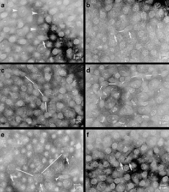
Fig. 4.2
Photographs of the cells on the basal membrane of whole mounts of seminiferous
tubules of a Chinese hamster, stained with hematoxylin staining the nuclei of the germ cells and
Sertoli cells. Sertoli cell nuclei can be recognized by their big nucleolus and the clumps of chro-
matin attached to them at either side. A few are indicated in (
a
) (
arrowheads
). As the tubule is a
three-dimensional structure, not all nuclei in an area will be in focus as they can be at a slightly
different level from the basal membrane. (
a
) A
s
spermatogonium (
arrow
). (
b
) A
pr
spermatogonia
(
arrows
). A
line
has been drawn between the cells of the pair. (
c
) A chain of four A
al
spermatogo-
nia. (
d
) A chain of nine A
al
spermatogonia that are part of a chain that continues beyond the area
of the photograph, consisting of in total 16 cells. (
e
) Three A1 spermatogonia (
arrows
) with rep-
resentative internuclear distances that are larger than those between the cells belonging to a clone
of A
pr
or A
al
spermatogonia. In this area there is also an A
s
spermatogonium (
arrowhead
). (
f
) Two
A spermatogonia close together that technically form a pair as their internuclear distance is less
than 25 mm. However, one of the nuclei is bigger than the other and has an elongated shape while
the other has a more oval nucleus. This likely is a false pair, i.e., two A
s
spermatogonia that have
stayed together. Bar = 8 mm
a phenomenon called the wave (Perey et al.
1961
). When one follows the wave
of spermatogenesis along a seminiferous tubule, the stages of the epithelial cycle
pass by and also one can follow the subsequent phases of the cell cycle of the
A1, A2, A3, A4, In, and B spermatogonia (Fig.
4.3
; Lok and de Rooij
1983a
).
For example, when a certain area contains A3 spermatogonia in G1 phase of the
Search WWH ::

Custom Search