Biomedical Engineering Reference
In-Depth Information
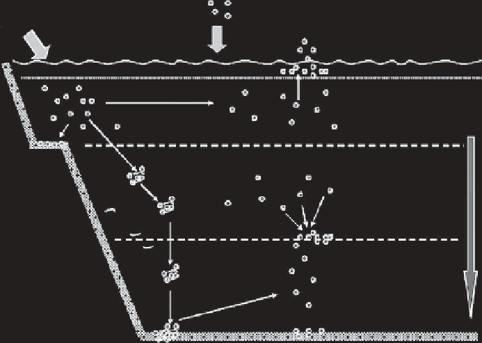
Particulate and organic matter
from coastal runoffs
Atmospheric
inputs
Formation of aerosol,
risk to seabirds, and mammals
To xicity to embryos and plankton
Concentration of NPs in the surface Microlayer
Changes in temperature
ionic strength, and natural
organic matter with depth
Dilution and transport
to open ocean
To xicity to pelagic species
Aggregation
Coastal
sediments
Accumulation of NPs
or aggregates at interfaces
Precipitation to
ocean floor
Mobilization of NPs
by microbes
To xicity to benthos
Ocean floor
FIGURE 1.12
Schematic diagram outlining the possible fate of nanoparticles (NPs) in the marine envi-
ronment and the organisms at risk of exposure. (Reprinted with permission from Klaine, S. J. et al. 2009.
Environmental Toxicology and Chemistry
27(9): 1825 -1851.)
1.6.2 N
aNotoxIcIty
IN
the
B
ody
NMs effect the human body at multiple levels, broadly differentiated into molecular, cellular, and
organular. The interaction of NMs with biomolecules, such as proteins and lipids, is multivari-
ate and complex. The nano-biointeractions of NMs with the physiological environment molecules
account for most of the toxicological effects induced by NMs.
At the cellular level, NMs may also cause mitochondrial injuries, enter the nucleus and dam-
age the DNA, depolarize cell membranes, and also physically damage the membranes by forming
nanosized holes. There are different methods by which NMs can interact with the cell membranes,
such as via hydrophobic forces, electrostatic forces, van der Waals forces, hydrogen bonding, or
receptor-ligand interactions. Once adsorbed on the surface of cells, NMs can be internalized by the
cells. Sometimes, the sharp edges of NMs erode the membrane's surface, leading to perforations.
The holes thus formed can act as direct entry points for NMs. Not only do they induce toxicity to the
organelles inside the cell, these perforations may also lead the leakage of intracellular fluid into the
surrounding medium and vice versa, thus inducing acute toxicity and possibly leading to cell death.
Cellular damage manifests itself at the organular level. As explained earlier, the production of
ROS can lead to oxidative stress in biological systems. Their production is believed to be the main
cause of induced toxicity in the blood, liver, spleen, kidneys, lungs, and any other organs with which
they come into contact. The resultant oxidative stress can produce proinflammatory cytokines, as it
is believed that ROS can affect the calcium-mediated signaling pathways within the cells.
1.6.2.1 Molecular Mechanisms of Nanomaterial Toxicity
Several different mechanisms have been proposed for the toxicity of NMs in the body (Figure 1.13).
The induction of oxidative stress via free radical formation is the prime molecular mechanism of
in
vivo
nanotoxicity (Lanone and Boczkowski 2006). These free radicals cause damage to biological
components through the oxidation of lipids, proteins, and DNA. As a consequence of this oxidative