Biomedical Engineering Reference
In-Depth Information
TABLE 18.2
Fine Description of the Synthetic Identity of the Dextran-Coated Nanoparticles Prepared
in Different Conditions
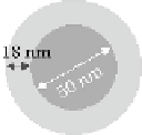
Nanoparticle A1
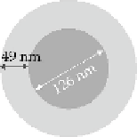
Nanoparticle R1
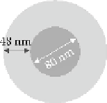
Nanoparticle R2
Size Characteristics
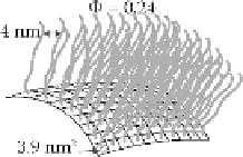
Arrangement of Dextran Chains in the Nanoparticle Corona
Fraction of the area of the core surface occupied by the anchorage of the polysaccharide chain:
F
Mesh size of the polysaccharide chains in the corona:
m
Maximal size of hydrophobic areas available on the nanoparticle core surface:
A
F
= 50%
a
F
= 20%
b
m
= 4.0 ± 0.2 nm
b
A
= 3.9 ± 0.6 nm²
b
F
= 7%
b
m
= 47 ± 2 nm
b
A
= 32 ± 5 nm²
b
Note:
See Table 18.1 for conditions of preparation. Φ gives the volume fraction occupied by dextran chains in the nanopar-
ticle corona.
a
Vauthier, C., Lindner, P., and Cabane, B., 2009. Configuration of bovine serum albumin adsorbed on polymer particles
with grafted dextran corona.
Coll. Surf. B: Biointerfaces
. 69:207-15.
b
Vauthier, C., Persson, B., Lindner, P., and Cabane, B. 2011. Protein adsorption and complement activation for di-block
copolymer nanoparticles.
Biomaterials
. 32:1646-56.
foreign bodies. This strategy may be used to target drugs to the immune system, but it hampers the
delivery of drugs to other organs and tissues. The approach used to escape the capture of drug car-
riers by macrophages is to mask the nanoparticle surface with hydrophilic polymers to modify the
composition and amount of adsorbed proteins when nanoparticles are injected into the blood. The
grafting of poly(ethylene glycol) (PEG) chains was a very efficient strategy to hamper the adsorption
of proteins onto the surface of nanomaterials used as drug carriers (Gref et al., 2000; Owens and
Peppas, 2006; Vonarbourg et al., 2006; Alexis et al., 2008; Li and Huang, 2008). It continues to be
actively studied, as the understanding of how the particle's design influences the adsorption of pro-
teins remains incomplete (Walkey et al., 2012; Walkey and Chang, 2012). Besides these works, we
have started investigating the interactions of proteins with the model nanoparticles, PACA coated
with polysaccharides, for which we could describe the synthetic identity of the hydrophilic corona.
Our studies was aimed to answer several questions:
•
Where were proteins adsorbed on the nanoparticle surface, assuming that they may remain
on the top of the hydrophilic corona, become entrapped within the hydrophilic corona, or
be adsorbed on the surface of the hydrophobic core?