Biomedical Engineering Reference
In-Depth Information
et al. 2009). Surface oxidation and other chemical/physical exchanges between the particle and GI
environment can produce unpredictable toxicities by ROS.
13.5.8 t
oxIcIty
of
c
hItosaN
Np
s
Nevertheless, while the chitosan bulk polymer is biocompatible, the polymer when presented as NPs
may not be quite as innocuous. Using lung and intestinal epithelial cell models, different mecha-
nisms of the cellular uptake and distribution of dissolved or nanoparticulate chitosan were estab-
lished (Huang et al. 2004; Loh et al. 2010; Ma and Lim 2003).
13.5.9 t
oxIcIty
of
g
old
Np
s
Pompa et al. investigated the effects of citrate-capped gold nanoparticles (Au NPs) upon ingestion by
the model system,
Drosophila melanogaster
. The significant
in vivo
toxicity of Au NPs was observed,
which elicited clear adverse effects in treated organisms, such as strong diminution of their life span
and fertility, DNA fragmentation, as well as a substantial overexpression of the stress proteins.
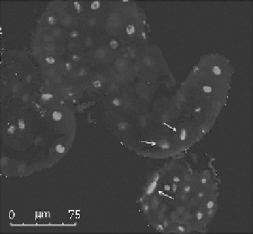
The GI tissue was further investigated to assess any occurrences of DNA damage in 15 nm Au
NP-treated individuals (100 pmol/L). For this purpose, a specific fluorescence kit based on termi-
nal deoxynucleotidyl transferase (TdT)-mediated terminal transferase dUTP nick-end-labeling
(TUNEL) was used. Importantly, among the numerous nuclei displaying no DNA damage, few cells
were observed in which DNA fragmentation was evident (Figure 13.7). Such DNA damage was found
to be distributed throughout the whole GI tissue with no apparent localization into specific regions.
Several midgut samples were analyzed with this technique and the observation of DNA nicks in
enterocytes was highly reproducible. On the contrary, in supernatant (SN)-treated samples as well
as in control flies, weak fluorescent signals related to DNA strand nicks were typically detected.
A quantitative analysis of the TUNEL assay revealed an occurrence of DNA damage of around
8% (8.15% ± 2.46%) in Au NP-treated flies, while in the control and SN-treated samples, DNA
fragmentation was <1% (0.84% ± 0.43%) (student's
t
-test
p
< 0.001). This experimental evidence
indicates a clear toxic effect of Au NPs on GI tissue. The localization of the 15 nm Au NPs in the
enterocytes, evidenced by TEM analyses (Figure 13.7), strongly suggests an indirect effect of the
nanomaterials in causing DNA damage. Further analyses are required to understand this point bet-
ter, but the observed fragmentation is very likely to be mediated by oxidative stress and/or related
to early-stage apoptosis (Pompa et al. 2011).
FIGURE 13.7
(
See color insert.
) Representative confocal microscopy image of
Drosophila
midgut in flies
treated with 15 nm Au NPs (100 pmol/L). Nuclei are stained with Hoechst 33 342 (blue) while cells contain-
ing DNA strand nicks are detected by TUNEL assay and fluorescent red (highlighted by the white arrows).