Biomedical Engineering Reference
In-Depth Information
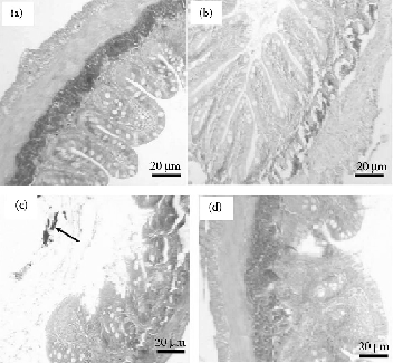
FIGURE 13.6
Histology of the intestine at the end of the experiment. (a) Fresh water control showing the
normal intestine, (b) solvent control, (c) intestine from a fish in 0.1 mg L
−1
SWCNT showing erosion of the
epithelium and precipitated SWCNT in the gut lumen (arrow), and (d) intestine from a fish in 0.5 mg L
−1
SWCNT showing fusion of the intestinal villi. Scale bar = 20 μm, sections were 8 μm thick and stained with
Mallory's trichrome. (Reproduced with permission from Smith, C.J., Shaw, B.J. and Handy, R.D. 2007.
Aquat
Toxicol
82:94 -109.)
Mammalian studies have raised concerns about the toxicity of CNTs; however, there are very
limited data on the ecotoxicity to aquatic life. Stock solutions of dispersed SWCNTs were prepared
using a combination of solvents (sodium dodecyl sulfate) and sonification. A semistatic test system
was used to expose rainbow trout to a solvent control, freshwater control, 0.1, 0.25, or 0.5 mg L
−1
SWCNTs for up to 10 days. Fish ingested water containing SWCNTs during exposure (presum-
ably stress-induced drinking) that resulted in precipitated SWCNTs in the gut lumen and intestinal
pathology (Smith et al. 2007).
The histology of the intestine is shown in Figure 13.6. Gross observations during dissection
showed clear black deposits in the gut lumen, which indicated that the fish had been drinking
the SWCNT-contaminated water. Subsequent histological examinations of the intestine showed
no effects from the control solvent, but demonstrated some clear intestinal pathology associated
with SWCNT exposure (Figure 13.6). All fishes from the 0.1 mg L
−1
SWCNT treatment showed
some areas of intestinal villi fusion, areas of inflammation and erosion, or total atrophy of the
mucosa. Precipitated SWCNT could be seen in the gut lumen (Figure 13.6c, arrow). The injuries
were observed to a lesser extent at 0.5 mg L
−1
(in four out of six of the fishes examined) and 1.0 mg
L
−1
SWCNT (half of the fishes examined). There was no evidence of major bleeding from the blood
vessels of the submucosa, but the tissue layer with the associated nerve plexus appeared more granu-
lar in the SWCNT-treated specimens than the controls (Smith et al. 2007).
13.5.5 t
oxIcIty
of
M
etal
Np
s
The zebrafish (
Danio rerio
) has become an important model species for the study of microbial
communities in vertebrate intestines (Rawls et al. 2004, 2007), and this model has also been