Biomedical Engineering Reference
In-Depth Information
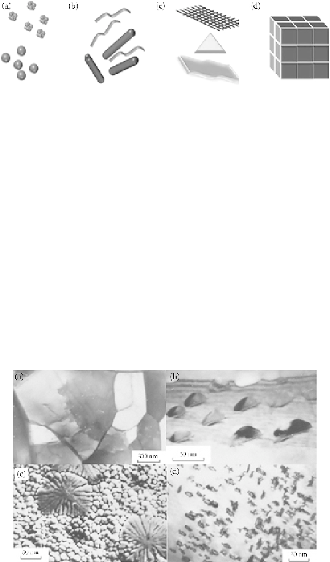
FIGURE 1.4
Classification of nanomaterials. (a) Zero-dimensional spheres and clusters. (b) One-dimensional
nanofibers, wires, and rods. (c) Two-dimensional films, plates, and networks. (d) Three-dimensional nanoma-
terials. (Reprinted with permission from Alagarasi, A. 2011.
Introduction to Nanomaterial
. National Centre
for Catalysis Research.)
NMs can be classified based on their phase composition properties, such as single-phase solids
(crystalline, amorphous particles, layers, etc.), multiphase solids (matrix composites, coated parti-
cles, etc.), and multiphase systems (colloids, aerogels, ferrofluids, etc.). Based on their methods of
manufacturing, NMs can be classified into three different categories: gas-phase reactions (flame
synthesis, condensation, etc.), liquid-phase reactions (sol-gel, precipitation, hydrothermal process-
ing, etc.), and mechanical procedures (ball milling, plastic deformation, etc.) (Wolfgang 2004). Based
on their structural properties, NMs can be classified into two parts: (1) nanocrystalline materials,
such as crystals, generally consisting of crystallite with at least one dimension in a nanometer size,
and (2) nanostructured materials, such as dislocation fragments, clusters, quasicrystals, micropores,
subgrains, and segregations. Nanofragmented materials, composed of dislocation fragments or sub-
grains whose size is less than 100 nm (Figure 1.5a), normally consist of metals and alloys subjected to
megaplastic deformations. Nonporous materials mainly exhibit a high volume density of nanopores
less than 100 nm situated on the conventional grain body or along their boundaries (Figure 1.5b).
Nanodendrites are materials mainly consisting dendrite solidification products in the form of degen-
erate dendrite nanodendrites, such as dendrite cells, and become visible upon the rapid solidification
FIGURE 1.5
Nanostructured materials. (a) Structure of nanofragmented material. Melts quenched FeSi
alloy, TEM. (b) Structure of nonporous materials. Nanopores are located at grain boundaries in a polycrystal-
line FeAl alloy produced by melt quenching, TEM. (c) Structure of nanodendrite materials. Dendrite nanocell
in side grain in a FeSi produced by melt quenching are visible, SEM. (d) Structure of nanodislocation materi-
als. Melt quench FeLi ally has high density of prismatic vacancy-type dislocation loop, TEM. (Reprinted with
permission from Glezer, A. M. 2011.
Russian Metallurgy (Metally)
4:263-269.)