Environmental Engineering Reference
In-Depth Information
250 nm
250 nm
(a)
3.0
3.0
2.0
2.0
1.0
1.0
3.0
µ
m
0
1.0
2.0
µ
m
0
1.0
2.0
3.0
nm
250
nm
200
0
0
-250
-200
µ
m
3.0
µ
m
0
1.0
2.0
3.0
0
1.0
2.0
(b)
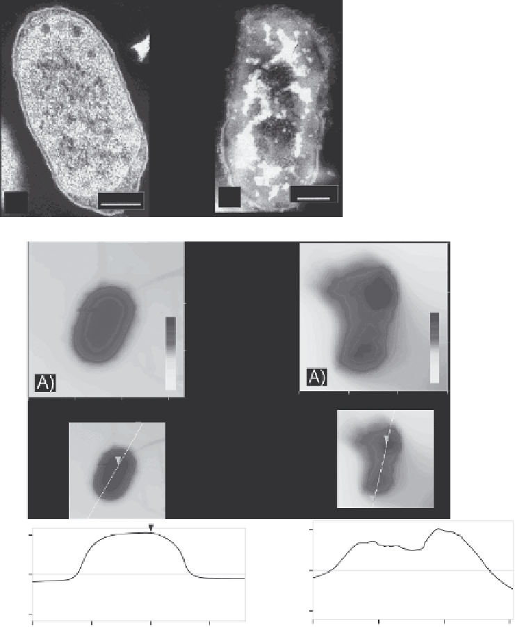
Figure 7.8
(a) TEM images of Escherichia coli cells Left: untreated. Right: treated with
halogenated MgO nanoparticles for 60 minutes. (b) Tapping mode AFM images of E. coli
cells with the corresponding cross sections below. Left: Untreated (z-height 0-920 nm).
Right: treated with halogenated MgO nanoparticles for 20 minutes (z-height 0-450 nm). Note
the changes in smoothness and height of the cell indicating damage to the E. coli cell enve-
lope upon nanoparticle treatment. (Reprinted with permission from P. K. Stoimenov, R. L.
Klinger, G. L. Marchin and K. J. Klabunde, Metal oxide nanoparticles as bactericidal agents,
Langmuir
,
18
, 6679-86. Copyright 2002 American Chemical Society.) (See colour plate
section for a colour representation)


Search WWH ::

Custom Search