Information Technology Reference
In-Depth Information
development of the hierarchy of visual areas probably proceeds from lower areas to
higher areas.
The repetitive patterns of V1 and V2 lead to the speculation that higher cortical
areas, like V4, IT, or MT contain even more complex interwoven feature maps. The
presence of many different features within a small cortical patch that belong to the
same image location has the clear advantage that they can interact with minimal wire
length. Since in the cortex long-range connections are costly, this is such a strong
advantage that the proximity of neurons almost always implies that they interact.
2.3 Layers
The cortical sheet, as well as other subcortical areas, is organized in layers. These
layers contain different types of neurons and have a characteristic connectivity. The
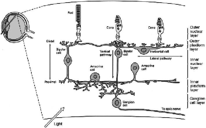
best studied example is the layered structure of the retina, illustrated in Figure 2.8.
The retina consists of three layers that contain cell bodies. The outer nuclear
layer contains the photosensitive rods and cones. The inner nuclear layer consists
of horizontal cells, bipolar cells, and amacrine cells. The ganglion cells are located
in the third layer. Two plexiform layers separate the nuclear layers. They contain
dendrites connecting the cells.
Information flows vertically from the photoreceptors via the bipolar cells to the
ganglion cells. Two types of bipolar cells exist that are either excited or inhibited by
the neurotransmitters released from the photoreceptors. They correspond to on/off
centers of receptive fields.
Fig. 2.8.
Retina. Spatiotemporal compression of information by lateral and vertical interac-
tions of neurons that are arranged in layers (adapted from [117]).
Search WWH ::

Custom Search