Information Technology Reference
In-Depth Information
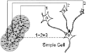
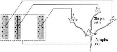
Fig. 2.2.
Simple and complex cells. According to Hubel and Wiesel [105] simple cells com-
bine the outputs of aligned concentric LGN cells. They respond to oriented stimuli and are
phase sensitive. The outputs of several simple cells that have the same orientation, but dif-
ferent phases are combined by a complex cell, which shows a phase invariant response to
oriented edges (adapted from [117]).
These cells send action potentials to a thalamic region, called lateral geniculate
nucleus (LGN). Different types of retinal ganglion cells represent different aspects
of a retinal image patch, the receptive field. Magnocellular (M) cells have a rela-
tively large receptive field and respond transiently to low-contrast stimuli and mo-
tion. On the other hand, parvocellular (P) ganglion cells show a sustained response
to color contrast and high-contrast black-and-white detail.
The optical nerve leaves the eye at the blind spot and splits into two parts at
the optical chiasma. Axons from both eyes that carry information about the same
hemisphere of the image are routed to the contralateral LGN, as can be seen in
Figure 2.1(b). In the LGN, the axons from both eyes terminate in different lay-
ers. Separation of P-cells and M-cells is maintained as well. The LGN cells have
center-surround receptive fields, and are thus sensitive to spatiotemporal contrast.
The topographic arrangement of the ganglion receptive fields is maintained in the
LGN. Hence, each layer contains a complete retinal map. Interestingly, about 75%
of the inputs to the LGN do not come from the retina, but originate in the cortex and
the brain stem. These top-down connections may be involved in generating attention
by modulating the LGN response.
From the LGN, the visual pathway proceeds to the primary visual cortex (V1).
Here, visual stimuli are represented in terms of locally oriented receptive fields.
Simple cells have a linear Gabor-like [79] response. According to Hubel and
Wiesel [105], they combine the outputs of several aligned concentric LGN cells (see
Fig. 2.2(a)). Complex cells show a phase-invariant response that may be computed
from the outputs of adjacent simple cells, as shown in Figure 2.2(b). In addition to
the orientation of edges, color information is also represented in V1 blobs. As in
the LGN, the V1 representation is still retinotopic - information from neighboring
image patches is processed at adjacent cortical locations. The topographic mapping
is nonlinear. It enlarges the fovea and assigns fewer resources to the processing of
peripheral stimuli.
Area V2 is located next to V1. It receives input from V1 and projects back to it.
V2 cells are also sensitive to orientation, but have larger receptive fields than those
in V1. A variety of hyper-complex cells exists in V2. They detect line endings,
Search WWH ::

Custom Search