Agriculture Reference
In-Depth Information
compartments were affected by the exposure to toxic agents leading to cellular inviability. In
these cases, damaged cells are expelled towards intestinal lumen.
Samples of landfarming and sewage sludge presented genotoxic action, evidenced by the
occurrence of nucleus fragmentation in the principal epithelial cells, karyolysis in the
nucleus of the cells of the fat body layer (Souza & Fontanetti, 2011) and loss of integrity of
the nuclear envelope of hepatic cells and cells of the “fat body” layer of the midgut (Nogarol
& Fontanetti, 2011).
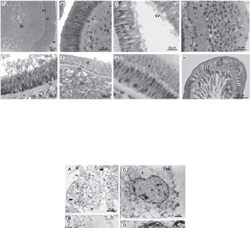
Fig. 4. Midgut of the diplopod
R. padbergi
stained with Hematoxylin-Eosin. Unexposed
animals (A; B); Animals exposed to sewage sludge (C-H). secretion vesicles (C); haemocytes
aglomeration (D); epithelium renewal (E); increase of cytoplasmatic granules in “fat body
layer” (F); cytoplasmatic vacuolization (G); volume reduction of the cells in “fat body” layer
of midgut (H). e=epithelium; m= muscle layer; fb= “fat body” layer; h= haemocytes; v=
vacuole; sv= secretion vesicle; * dilatation of intercellular space (Photos: Larissa Rosa
Nogarol; Raphael Bastão de Souza and Tatiana da Silva Souza)
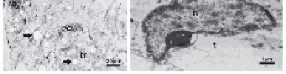
Fig. 5. Perivisceral fat body of the diplopod
R. padbergi
stained with Hematoxylin-Eosin (A;
B) and submitted to TEM routine (C; D). Unexposed animal (A); Animal exposed to sewage
sludge (B-D). Loss of cell limit and increase of spherocrystal (B); Cytoplasmatic
vacuolization and loss of cell membrane integrity (C); Nucleus deformation (D). t=
trophocyte; tr = tracheoles; o= oenocyte; m= mitochondria; n= nucleus; v= vacuole; arrows=
spherocrystals; *= loss of cell membrane integrity. (Photos: Raphael Bastão de Souza and
Larissa Rosa Nogarol)