Biology Reference
In-Depth Information
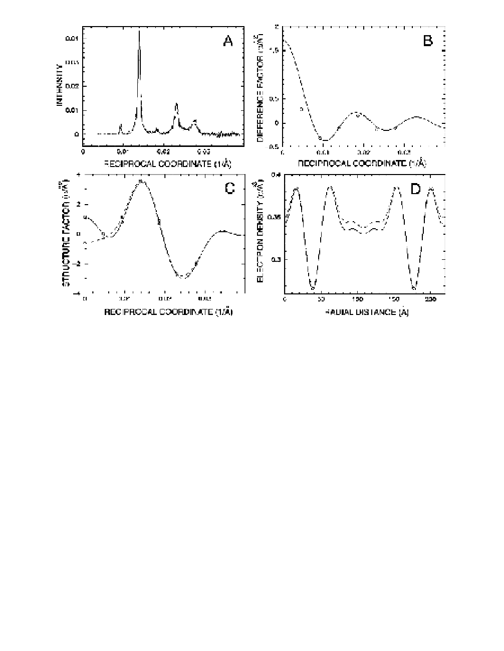
Fig. 3.
(A) Densitometer tracings of X-ray diffraction patterns after background
subtraction for mouse sciatic nerve treated in buffered medium with fluid electron
density of 0.3347 e/Å
3
at ionic strength 0.06 and pH 6.0 (solid line) and for the nerve
treated in the same buffered medium containing 10% glycerol with fluid electron
density 0.3474 e/Å
3
(dashed line). The myelin period for mouse sciatic nerve in the two
different media is 216 Å. (B) Difference Fourier transform (control structure factors
minus those from glycerol treated myelin) between the structure factors with different
fluid electron densities. The continuous curve is calculated and the circles are from
the observed structure factors. Using 2.0 for the scale parameter and 136 Å for the
exclusion length give 43% as the best goodness-of-fit. (C) The continuous Fourier
transform and the structure amplitudes on an absolute scale (e/Å
2
) in control medium
(solid line and circle) and in 10% glycerol solution (dashed line and triangle).
(D) Absolute electron density profiles of the membrane pair for the control (solid line)
and the glycerol-treated nerve (dashed line). The difference in the two density profiles
is at the extracellular space. The average electron density within the exclusion length
is 0.343 e/Å
3
. The cytoplasmic separation (32 Å), the distance between the polar
head groups (46 Å), and the extracellular separation (92 Å) are measured from the
profile.

Search WWH ::

Custom Search