Biology Reference
In-Depth Information
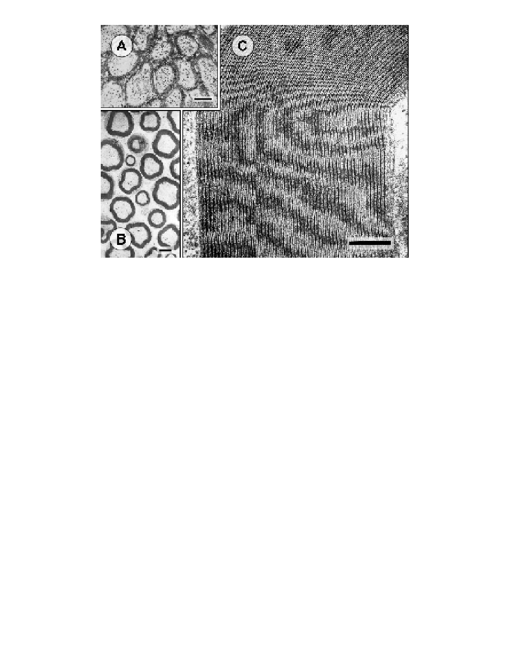
Fig. 1.
The ultrastructure of nerve myelin is revealed by electron micrographs. Tissues
of mice were prepared by chemical stabilization using glutaraldehyde, then dehydrated,
and embedded in plastic (Kirschner, Hollingshead, 1980). The plastic blocks containing
tissue were cut into thin-sections ∼800 Å thick, stained by heavy metals (lead or uranyl
salts), and observed at high magnification in the electron microscope. The images are from
tissue that had been sectioned at right angles to the long axis of the nerve, and therefore
the myelin appears at low magnification as darkly-stained rings. (A) Optic nerve. Note that
the different myelinated fibers abut one another. The dense bands that frequently traverse
the width of the myelin sheaths are specialized junctions that are unique to central nerv-
ous system myelin (Kosaras, Kirschner, 1990). Scale bar: 1 µm. (B) Sciatic nerve. The
individual myelinated fibers are separated from one another by collagen fibers. Scale bar:
3 µm. (C) High magnification view of a portion of one myelin sheath from sciatic nerve.
The densely-staining interface between cytoplasmic faces of the membranes (“major dense
line”) are clearly distinguished from the more lightly-staining surfaces of the extracellular
faces of the membranes (“intraperiod line”). The most electron lucent feature in the peri-
odic array of membrane pairs is the center of the membrane bilayers. Scale bar: 0.2 µm.
(The micrographs were obtained by Dr Béla Kosaras, Dr Allen L Ganser, and Ms Carol J
Hollingshead, respectively, in the laboratory of Dr DA Kirschner.)
multilamellar structure between nodes of Ranvier, as shown by the com-
plementary structural techniques of electron microscopy (Fig. 1) and X-ray
diffraction (Fig. 2) (Schmitt
et al
., 1941; Robertson, 1958). The sheath
results from the spiral wrapping of the plasma membranes of Schwann

Search WWH ::

Custom Search