Biology Reference
In-Depth Information
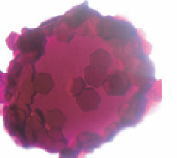
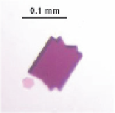
Fig. 6.
A simplified representation of crystallization from vesicles obtained from
2-D crystalline native purple membranes. The authors of this method suggest that BR
crystals are formed by fusion of the vesicles. (The schematic diagram on the left-hand side
was modified from Takeda
et al
. (1998)). The crystals grown in our laboratories are also
shown (Golubev
et al
., unpublished).
that one can obtain different types of BR crystals (Fig. 6). However, at
present, no other membrane proteins have been crystallized by this
method. Nevertheless, it is not yet clear whether this approach is limited
to some specific cases, like BR, or if it has a more general application.
Unfortunately, as in the two other cases mentioned above, this system of
crystallization has not yet been sufficiently characterized.
It is worth noting that there is an additional side of biological signifi-
cance of these studies. For instance, the construction of artificial highly
curved aggregates could help to understand more deeply the process of
vesicle formation in living organisms. Indeed, well-known examples of the
important roles of protein-containing spherical vesicles in eukariotic cells
are: the clathrin coated vesicles that realize endocytosis of ligands bound
to the cell surface; the coatomer coated vesicles that transport proteins to
and from the Golgi complex; and synaptic vesicles in the nerve cells that
transfer the neurotransmitters from the synaptic cell into the synaptic cleft.
In addition, one should not forget proteins assembled in viral capsids.


Search WWH ::

Custom Search