Biology Reference
In-Depth Information
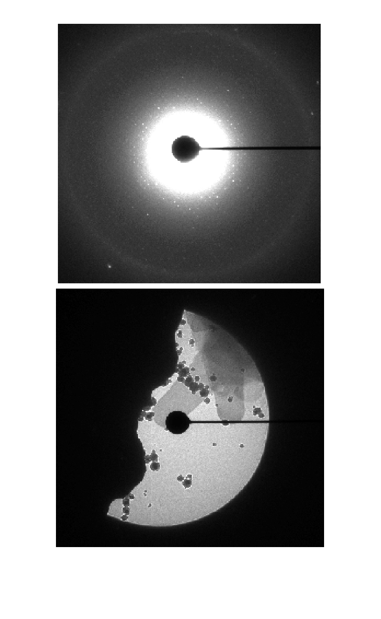
(A)
(B)
Fig. 3.
Electron diffraction pattern from a 2D crystal. The pattern in (A) was recorded
from the crystal in (B), which is partly hidden by the beam stop used to protect the
CCD camera from the primary beam. The black dots in the image originate from ice
contamination.


Search WWH ::

Custom Search