Biomedical Engineering Reference
In-Depth Information
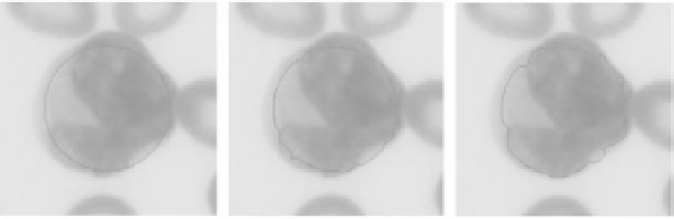
Figure 1.
The image segmentation result of a Follicular Center Cell Lymphoma (FCC)
applying traditional deformable model. The left panel shows the initial position, the center
panel shows one snapshot of the evolving contours, and the right panel shows the final
segmentation results after 150 iterations. See attached CD for color version.
and
A
and
x
spatially defined as
lqp
00
qlqp
0
pq l qp
0
pq l q
00
pq l
x
i−
2
x
i−
1
x
i
x
i
+1
x
i
+2
y
i−
2
y
i−
1
y
i
y
i
+1
y
i
+2
A
=
x
=
y
=
,
,
,
and
p
=
β
,
q
=
−
−
4
β
, and
l
=2
α
+6
β
.
The limitations of the traditional deformablemodel are its small capture region
and sensitivity to initial (starting) position. Figure 1 shows the performance of
applying the traditional deformable model on segmenting a Follicular Center Cell
Lymphoma (FCC). It can be seen that if the initial position is not sufficiently close
to the object boundary, the traditional deformable model cannot segment the nuclei
accurately. (Please note that all segmentation results are obtained after applying
the color gradient described in [17], whereas using the traditional gradient[4] does
not result in satisfactory convergence to the nuclear boundary.)
α
3.2. Balloon Deformable Model
In order to resolve the difficulties with the small capture region of the tradi-
tional deformable model, Cohen and Cohen [13] proposed a new external force
that could enlarge the scope of the capture zone of the original model. Instead of
defining
E
ext
as the negative gradient of the image, they define
E
ext
=
−∇
P
(
x
)
,
(22)
where
P
(
s
) is the potential function calculated using a Euclidean (or Chamfer)
distance map. Let
d
(
x
) be the distance between a point
x
and the nearest edge