Biomedical Engineering Reference
In-Depth Information
(a)
(b)
(c)
(d)
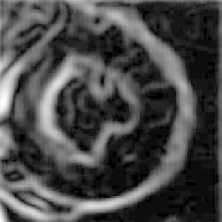
Figure 11.
Illustration for performance of gradient vector flow (GVF) snake in segmentation
of left ventricle from an MR image of heart: (a) original MR image showing left ventricle;
(b) gradient map derived from (a); (c) vector flowfield of the gradient map; (d) segmentation
result using GVF snake. Reprinted with permission from [25]. Copyright c
1998, IEEE.
The basic nature of this force field is such that deformation near the nonho-
mogeneous regions of the image is governed by the gradient function, while in
the homogeneous region the first term dominates. This results in a flow vector
that converges into the edges of the image. Figure 11 illustrates the performance
of gradient vector flow-based active contour model in segmentation of the left
ventricle from MR images of the heart.
Anatomical structures varywidely in shape, size, and geometry. Segmentation
of this wide range of anatomical structures requires highly deformable models to