Biomedical Engineering Reference
In-Depth Information
(a)
(b)
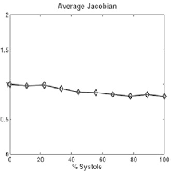
Figure 27.
Metrics for the left ventricle of a patient with a history of myocardial infarction
illustrated by (a) the Jacobian for the Lagrangian fit that registers
R→R
t
, and (b) the
residuals for fitting
R
t
→R
.
shortening during left-ventricular contraction results in the negative strain values
in the circumferential direction, while compression in the longitudinal direction
results in negative longitudinal strains. These results are comparable with other
relevant work ([2, 33]). Qualitative strain results for one dog study are shown
in Figure 19. Strain maps are reconstructed on the midventricular surfaces of
the biventricular model. The RV surface (
u
RV
=0
.
5) exhibits circumferential
shortening (Figure 19b), consistent with the segmental shortening seen in [5, 6, 7].
Regional peak principal strains (
E
3
) are given in Table 5 and are consistent with
those given in [4].
6.5.2. Human data
The general strain patterns seen in the canine data, i.e., positive radial strains
and negative circumferential and longitudinal strains, are also seen in the normal
human volunteer data (Figure 28). These strain patterns have similar physiological
interpretations. Comparison with Table 2 from Moore et al.'s work [33] demon-
strates that the three strain values in the LV are consistent with the range of strain
values in previous findings. The Eulerian strains depicted in Figure 29 are not as
smooth as their Lagrangian counterparts. This is to be expected since temporal
lofting is not performed for the Eulerian fits.
The strains from the clinical data (Figure 30) are more difficult to interpret.
While some of the regional plots are similar to their counterparts of the normal
human data, some of the plots demonstrate disparate behavior. Additional insight