Biomedical Engineering Reference
In-Depth Information
Table 5.
Peak Systolic Principal Strains (
E
3
) for
Six Right-Ventricular Regions of Dog 3
Base
Mid-Cavity
Anterior
−
0
.
12
±
0
.
004
−
0
.
22
±
0
.
018
Mid
−
0
.
10
±
0
.
001
−
0
.
16
±
0
.
008
Inferior
−
0
.
20
±
0
.
007
−
0
.
18
±
0
.
007
6.4. In Vivo Human Data
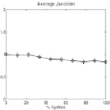
Also included in this analysis is an illustration of the application of our method
to systolic human datasets. One dataset is from a normal human volunteer (Fig-
ures 26, 28, and 29) and one dataset is from a patient with a history of myocardial
infarction (Figures 27 and 30). For each human study we have included the aver-
age Jacobian over 12 left-ventricular regions as well as the weighted least-squares
residuals of the fitting algorithm. For each case, we also illustrate the left ventric-
ular radial, circumferential, and longitudinal strains.
6.5. Interpretation of Results
6.5.1. Canine data
Normal LV strains, i.e., average radial, circumferential, and longitudinal
strains, are given in Figures 20 and 21. The radial strains remain positive for most
of the twelve regions, indicative of the systolic thickening of the left ventricle.
Both the circumferential and longitudinal strains are negative.
Circumferential
(a)
(b)
(c)
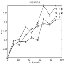
Figure 26.
Metrics for the left ventricle of a normal human volunteer illustrated by (a) the
Jacobian for the Lagrangian fit, (b) the Jacobian for the Eulerian fit that registers
R
t
→R
,
and (c) the residuals for fitting
R
t
→R
.